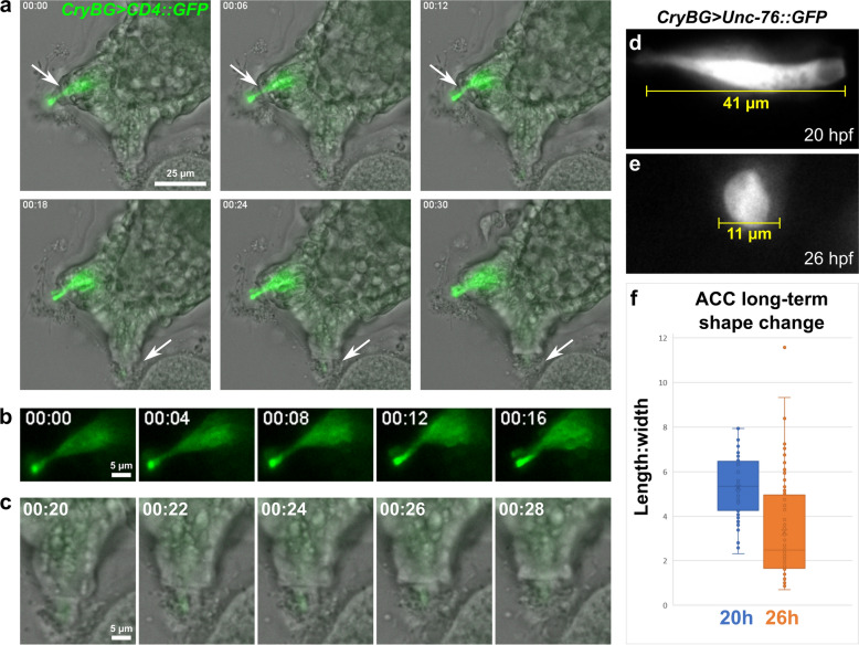
Fig. 4.
The contractility and shape change of ACCs during papilla retraction. a Still images captured from live time-lapse video (one animal recorded, see Additional file 4: Video S1) of attached larva ~ 21 h post-fertilization (hpf). ACCs marked by CryBG > CD4::GFP expression. Right dorsal papilla (top arrows) retracts from 6 to 12 min timepoints, while unlabeled left dorsal papilla (bottom arrows) retracts from 18 to 24 min. b Same GFP-labeled papilla in a, but only GFP channel shown, revealing ACC contraction. c Same unlabeled papilla in a, showing the finger-like apical protrusion of the ACC retracting into papilla. f Representative image of an ACC labeled by CryBG > Unc-76::GFP at 20 hpf, indicating typical length at this stage prior to settlement. e representative image of labeled ACC at 26 hpf, indicating extreme rounded shape observed in many larvae at this stage, after settlement and metamorphosis begins. f Quantification of length:width ratio of ACCs sampled from 20 vs. 26 hpf larvae (n = 54 for 20 hpf, n = 84 for 26 hpf)

