Fig. 1.
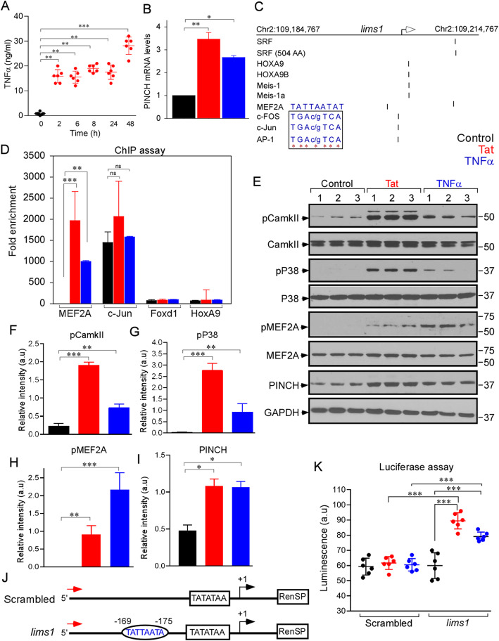
PINCH is transcriptionally regulated by MEF2A under inflammatory conditions. a Quantification of TNFα production by neurons exposed to Tat at different time points. b Quantification of PINCH mRNA levels in neurons untreated or exposed to Tat or TNFα for 48 h. c Bioinformatic analysis of lims1/pinch promoter sequence predicted a conserved putative binding site for different transcription factors (TF). Inset: Sequences show conserved binding sites for MEF2A, Cc-FOS, Cc-Jun, and AP-1. d ChIP-assay was performed in neurons untreated or exposed to Tat or TNFα. Antibodies specific for MEF2A, c-Jun, Foxd1, and HoxA9 were used to immunoprecipitate the chromatin and the fold enrichment of lims1/pinch promoter relative to the matched input control was quantified by qPCR. e Representative Western blot of lysates from neurons untreated or exposed to Tat or TNFα and probed with antibodies specific for phospho-CamkII, CamkII, phospho-P38, P38, phospho-MEF2A, MEF2A, PINCH and GAPDH. f-i Quantification of relative protein abundance of phospho-CamkII/CamkII (f), phospho-P38/P38 (g), phospho-MEF2A/MEF2A (h), and PINCH/GAPDH (i) from (e). j Schematic representation of the lims1/pinch promoter-luciferase constructs. The MEF2A consensus response element at − 169-175 base pairs (bp) (TATTATA) is shown in the oval. k Neurons transfected with control and lims1/pinch luciferase constructs were untreated or exposed to Tat or TNFα for 48 h and luciferase activity was measured. Data represent mean ± SEM; *P < 0.05; **P < 0.01; ***P < 0.001; n = 3–5 (one-way ANOVA)

