Fig. 3.
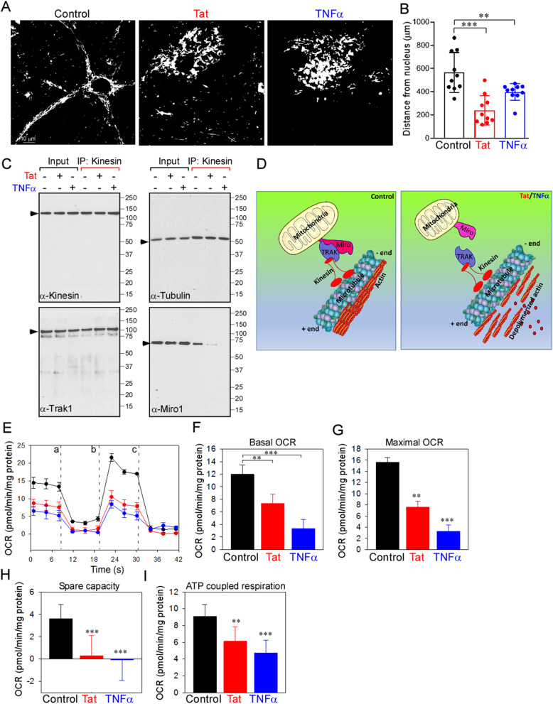
Increased PINCH expression and actin disassembly are accompanied by perinuclear localization of mitochondria. a Neurons untreated or exposed to Tat or TNFα for 48 h were monitored for cellular mitochondrial distribution using confocal microscopy. Neurons were stained with dihydrorhodamine (DHR123) and changes in mitochondrial distribution were observed. Representative confocal images show perinuclear localization of mitochondria in neurons exposed to Tat or TNFα. b Quantification of the distance (μm) of mitochondria from the nucleus. c Cell lysates from control, Tat- or TNFα-treated neurons were immunoprecipitated with antibodies against kinesin. Following immunoprecipitation, total cell lysates (input) and immunoprecipitated materials (IP) were subjected to Western analysis. Samples were probed with antibodies specific for Kinesin (top left), Tubulin (top right), Trak1 (bottom left), and Miro1 (bottom right). d Schematic representation shows the disruption of Kinesin-Trak-Miro complex in neurons exposed to Tat or TNFα. e-i Neurons were untreated or exposed to Tat or TNFα for 48 h and oxygen consumption rate (OCR) was measured. Untreated neurons were used as control. After measurement of baseline OCR, neurons were sequentially exposed to oligomycin (a), carbonyl cyanide-4-(trifluoromethoxy)phenylhydrazone (FCCP) (b), rotenone and antimycin A (c). Representative traces of OCR in control and neurons exposed to Tat/TNFα (e). Quantification basal (f), maximal (g), spare capacity (h), and ATP coupled respiration (i) in control and neurons treated with Tat/TNFα. Data represent mean ± SEM; **P < 0.01; ***P < 0.001; n = 3–5 (one-way ANOVA)

