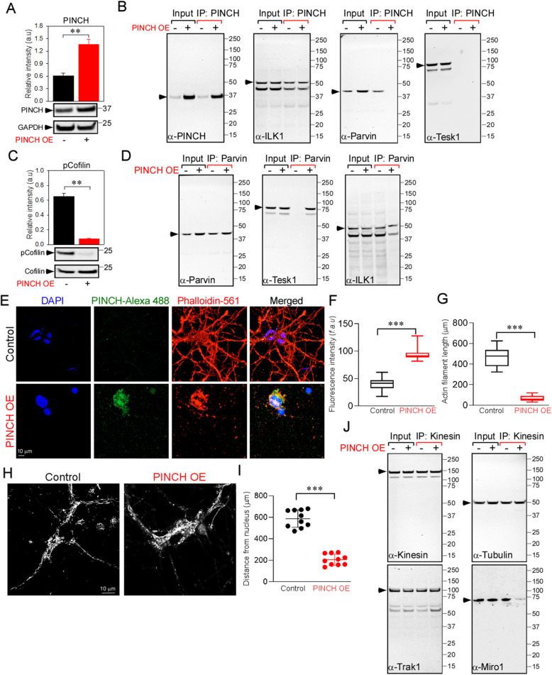
Fig. 4.
Exogenous expression of PINCH mimics the effects of Tat or TNFα exposure in neurons. a Quantification of relative protein abundance (PINCH/GAPDH) and representative Western blots for lysates from neurons with or without exogenous PINCH expression. b Cell lysates from control and PINCH-overexpressing (OE) neurons were immunoprecipitated with antibody specific for PINCH. Following immunoprecipitation, total cell lysates (input) and immunoprecipitated materials (IP) were subjected to Western analysis. Samples were probed with antibodies specific for PINCH, ILK1, α-Parvin, and TESK1. c Quantification of relative protein abundance (phospho-Cofilin/Cofilin) and representative Western blots for lysates from neurons with or without exogenous PINCH OE. d Cell lysates from neurons with or without exogenous PINCH OE were immunoprecipitated with antibody specific for Parvin. Following immunoprecipitation, total cell lysates (input) and immunoprecipitated materials (IP) were subjected to Western blot analysis using antibodies specific for Parvin, TESK, and ILK1. e Changes in cellular actin cytoarchitecture were observed using confocal microscopy in neurons with or without exogenous PINCH OE. Neurons were fixed, permeabilized and stained with anti-PINCH and phalloidin, the dye that stains actin filaments. Representative confocal images show depolymerization of actin in neurons exogenously expressing PINCH. (f and g) Quantification of PINCH fluorescence (f) and actin filament length (g). h Cellular mitochondrial distribution was observed in neurons with or without PINCH expression using confocal microscopy. Neurons were stained with dihydrorhodamine (DHR123) and changes in mitochondrial distribution were observed. Representative confocal images show perinuclear localization of mitochondria in neurons exogenously expressing PINCH. (i) Quantification of the distance of mitochondria from the nucleus. j Cell lysates from neurons with or without exogenous PINCH expression were immunoprecipitated with antibodies specific for kinesin. Following immunoprecipitation, total cell lysates (input) and immunoprecipitated materials (IP) were subjected to Western blot using antibodies specific for kinesin, Tubulin, Trak1, and Miro1. Data represent mean ± SEM; ***P < 0.001; n = 3–5 (one-way ANOVA)

