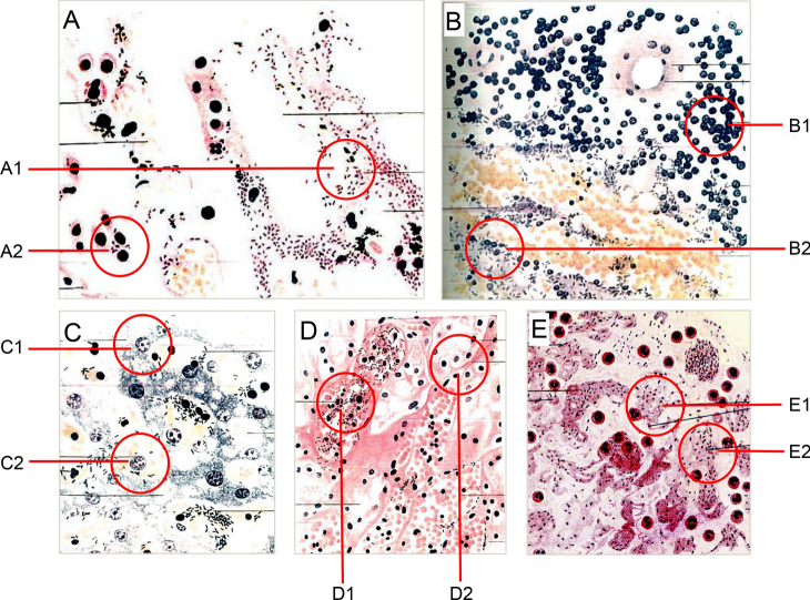
Fig. 1.
Pathological and bacteriological findings of infected organs and sputum of patients with pneumonic plague. A Lung section (300×, Giemsa’s stain). A1 Collapsed alveolar space. A2 Large epithelial cells containing ingested plague bacilli and carbon pigment. B Spleen section (200×, Giemsa’s stain). B1 Swollen and vacuolated lymphocytes. B2 Swollen trabecula having an enormous number of bacilli. C Liver section (10,000×, Giemsa’s stain). C1 Granular, vacuolated, and disintegrating liver cells. C2 Phagocytes in the walls of the portal capillary containing plague bacilli. D Section from the boundary zone of the kidney (250×, Giemsa’s stain). D1 Congested vessels, one of which contained abundant plague bacilli. D2 Swollen basement membranes of tubules and capillaries. E Detection of Yersinia pestis in the sputum (1000×, eosin-methylene blue). E1 A single Yersinia pestis cell. E2 Yersinia pestis in chains (A–D are from Wu 1914; E is from Wu 1926).

