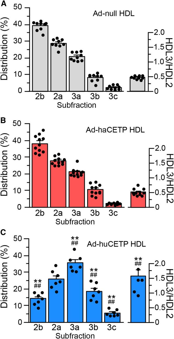
Fig. 4.
HDL particle size. HDL isolated from the indicated adenovirus group was fractionated by nondenaturing gradient gel electrophoresis. HDL protein was detected with Coomassie blue stain. A–C: HDL subfraction distribution. Values are mean ± SEM. See Fig. 2 for group sizes. **P < 0.01 versus Ad-null; ##P < 0.01 versus Ad-haCETP.

