Figure 7.

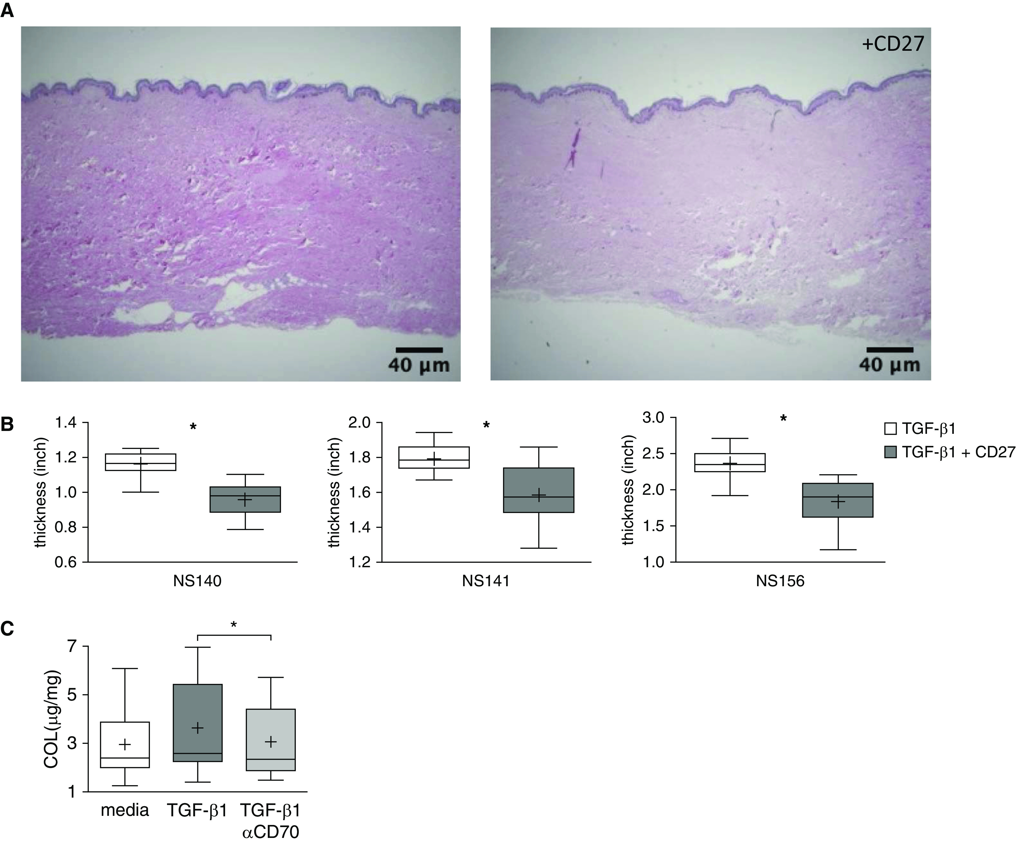
CD70 activation decreases ex vivo fibrosis. (A) Representative cross-sectional images of cultured human abdominal skin tissues injected with TGF-β1 (10 ng/skin sample), with or without concomitant injections of CD27 fusion protein (5 μg/skin sample) (hematoxylin and eosin stain). These preparations were harvested and imaged after treatment for 1 week. Scale bars, 40 μm. (B) Dermal thicknesses were quantified by averaging 19 random vertical measurements of histological cross-sections on each image. Each graph represents one set of these measures in each of the abdominal skin samples from three distinct individuals (NS140, NS141, and NS156). *P < 0.001. The lowest, second lowest, middle, second highest, and highest lines represent 10th, 25th, median, 75th, and 90th percentiles, respectively. Mean is denoted by (+). (C) Human foreskin punch biopsy samples from 20 distinct subjects were cultured in media supplemented with TGF-β1 (10 ng/ml), with or without anti-CD70 antibody (5 μg/ml), for 6 days. Hydroxyproline content, normalized to the dried weight of each skin specimen, was diminished in the specimens that received anti-CD70 treatment. *P = 0.003.
