
Figure 3.
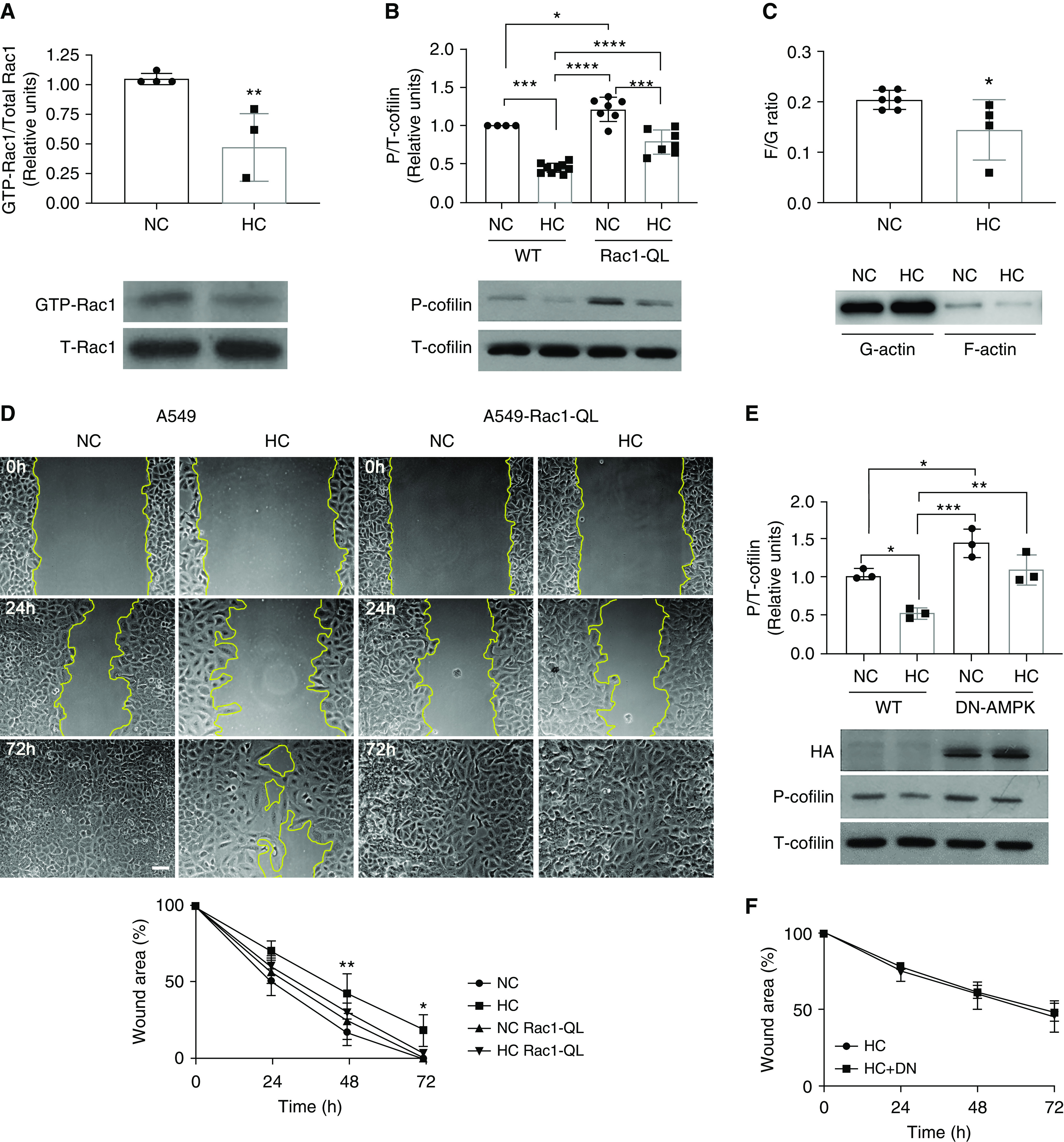
Hypercapnia impairs epithelial cell migration by inhibiting the Rac1/cofilin pathway via AMPK (AMP kinase). (A) Rac1 pull-down assay was performed in cell lysates from A549 cells exposed for 24 hours to NC or HC. Graph shows composite from different experiments (n = 4). Lower panel depicts a representative Western blot. Data were analyzed by unpaired t test. **P < 0.01. (B) A549-wild-type (WT) cells and A549-Rac1-QL cells were exposed for 24 hours to NC or HC, and Western blot against phospho-cofilin (P-cofilin) and total-cofilin (T-cofilin) was performed. Graph shows quantification of different experiments (n = 7–9). Lower panel depicts a representative Western blot. Data were analyzed by one-way ANOVA followed by Tukey multiple comparison test. *P < 0.05, ***P < 0.001, and ****P < 0.0001. (C) F- and G-actin were isolated using a commercially available kit from A549 cells exposed to NC or HC for 24 hours. Graph shows composite from different experiments (n = 4–6) of the F- to G-actin ratio. Lower panel depicts a representative Western blot. Data were analyzed by unpaired t test. *P < 0.05. (D) A549-WT and A549-Rac1-QL cells were placed for 24 hours in NC or HC before a scratch wound was made with a sterile pipette tip. Cells were maintained in NC or HC for the indicated times, and photographs were acquired. Graph shows wound area versus time (n = 4). Data were analyzed by two-way ANOVA followed by Sidak multiple comparison test. *P < 0.05 and **P < 0.01 compared with NC. Scale bar, 100 μm. (E) A549 cells were infected with adenovirus expressing DN-AMPK (dominant negative AMPK), exposed for 24 hours to NC or HC, and Western blot against P- and T-cofilin was performed. Graph shows quantification of different experiments (n = 3). Lower panel depicts a representative Western blot. HA (hemagglutinin) shows expression of the DN-AMPK. Data were analyzed by one-way ANOVA followed by Tukey multiple comparison test. *P < 0.05, **P < 0.01, and ***P < 0.001. (F) A549-WT and A549 cells infected with adenovirus expressing DN-AMPK were placed for 24 hours in NC or HC before a scratch wound was made with a sterile pipette tip. Cells were maintained in NC or HC for the indicated times, and photographs were acquired. Graph shows wound area versus time (n = 3). Data were analyzed by two-way ANOVA followed by Sidak multiple comparison test.