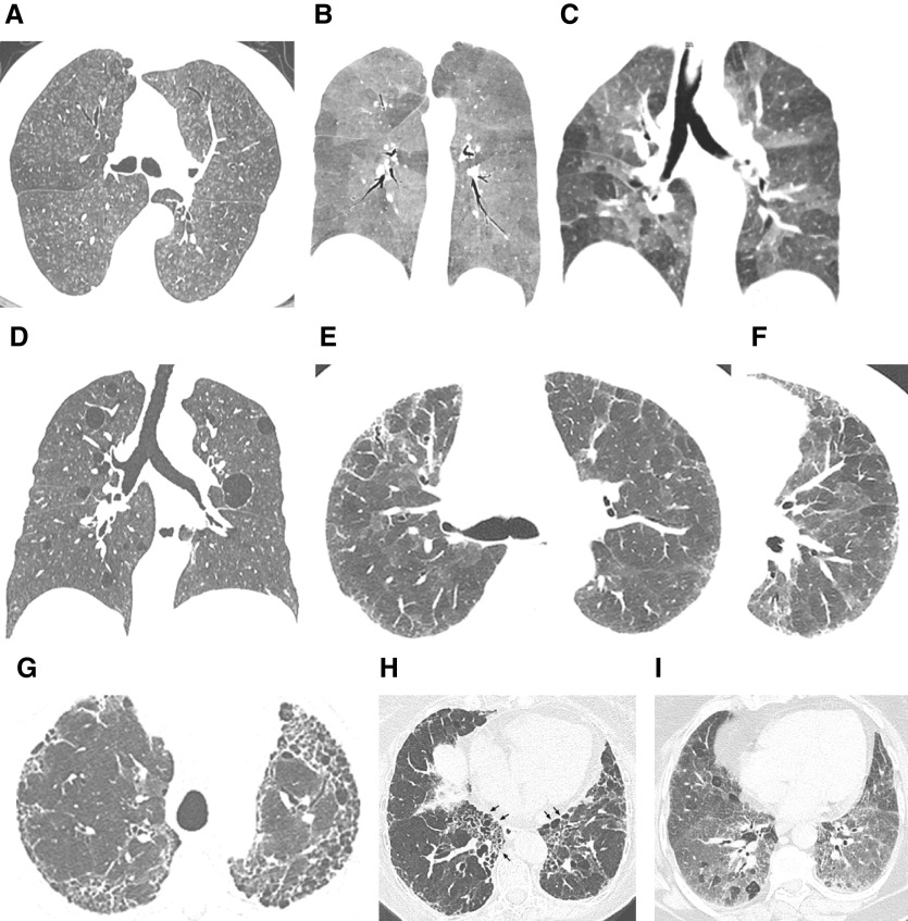
Figure 1.
“Typical hypersensitivity pneumonitis (HP)” and “compatible-with-HP” high-resolution computed tomography patterns. The nonfibrotic typical HP pattern is characterized by (A) centrilobular nodules, (B) mosaic attenuation on an inspiratory scan, and (C) air trapping on an expiratory scan. (D) The nonfibrotic compatible-with-HP pattern is exemplified by uniform and subtle ground-glass opacity and cysts. The fibrotic typical HP pattern consists of (E) coarse reticulation and minimal honeycombing in a random axial distribution with no zonal predominance in association with (F) small airway disease. The fibrotic compatible-with-HP pattern varies in the patterns and/or distribution of lung fibrosis (e.g., basal and subpleural predominance, [G] upper-lung-zone predominance, [H] central [or peribronchovascular] predominance [arrows], or [I] fibrotic ground-glass attenuation seen alone or in association with small airway disease). The fibrotic indeterminate-for-HP pattern includes the usual interstitial pneumonia pattern, nonspecific interstitial pneumonia pattern, organizing pneumonia–like pattern, or truly indeterminate findings.

