Figure 4.
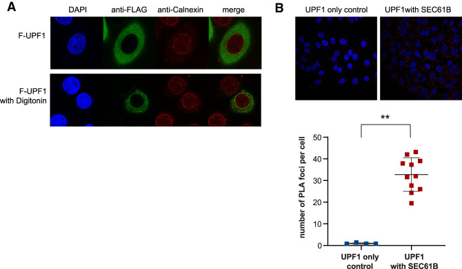
UPF1 localizes at the ER. (A) Immunofluorescence of HeLa cells transiently expressing Flag-tagged UPF1 (F-UPF1, in green) together with the ER marker Calnexin (in red). (Top) Cell nuclei were visualized by DAPI staining. UPF1 predominantly shows a diffused cytoplasmic localization. (Bottom) Partial cell permeabilization with digitonin revealed that a fraction of UPF1 is anchored at the ER membrane and colocalizes with calnexin. (B) UPF1 is localized in the close proximity of the SEC61B translocon component at the ER. Proximity ligation assay (PLA) using antibodies against endogenous UPF1 and SEC61B proteins generated a discrete signal (red spots), indicating that the proteins are <40 nm apart. The graph shows the quantification of PLA signal. Each point represents mean PLA count per cell in one captured frame. Significance was determined by two-tailed Mann-Whitney test. (**) P<0.005. Data showing the colocalization of NBAS and UPF2 with SEC61B are shown in Supplemental Figure S5.

