
Figure 2. Antitumor Activity In Vivo of iHAP1 versus PPZ in T-ALL Preclinical Models.
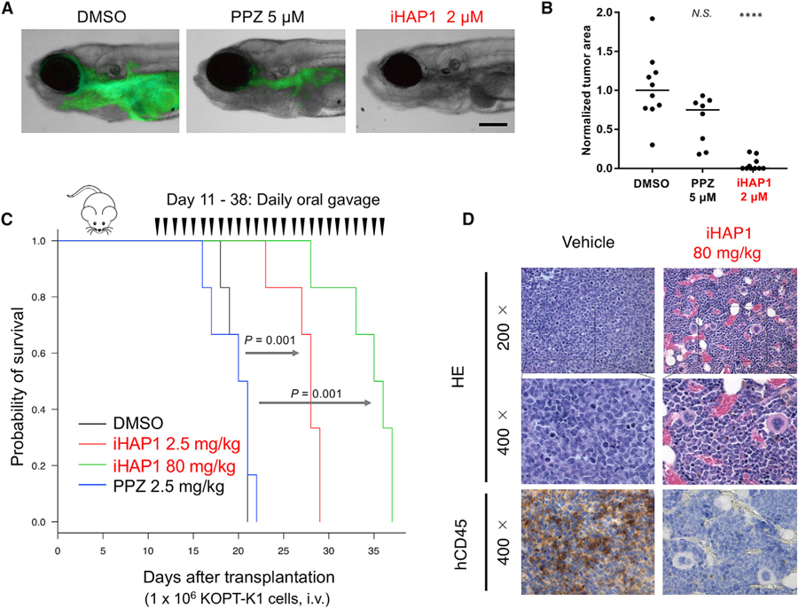
(A) Representative zebrafish embryos transplanted with GFP+ T-ALL cells isolated from Tg(rag2:Myc; rag2:EGFP) zebrafish and treated for 5 days with DMSO, 5 µM PPZ, or2 µM iHAP1. Scale bar, 0.1 mm.
(B) Quantification of GFP+ leukemic areas in treated zebrafish embryos. ****p < 0.0001 versus DMSO by two-tailed Welch’s t test; black bars indicate median values. N.S., not significant.
(C) Kaplan-Meier survival analysis of NSG mice xenotransplanted with 106 KOPT-K1 human T-ALL cells (day 1) and treated from day 11 as indicated on the plot. Six mice were tested in each cohort; p values were determined with a log rank test.
(D) Representative results of hematoxylin and eosin (H&E) and immunohistochemistry (IHC) staining of sections of femora from xenotransplanted NSG mice on day 18 post-xenotransplant and 24 h after mice received the seventh daily dose of drug p.o. (vehicle or iHAP1, 80 mg/kg). Engraftment and expansion of transplanted KOPT-K1 cells were detected by anti-human CD45 antibody.