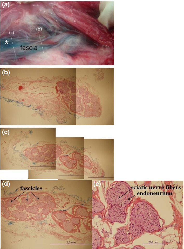
FIGURE 2.

The histopathological study of the sciatic nerve by the H&E staining. (a) post‐staining perineural space of sciatic nerve (*) (b) cross section of the sciatic nerve at the site of ‘before‐bifurcation’ (c) cross section of the sciatic nerve at the site of ‘post‐bifurcation’ (d) magnification of figure c in which methylene blue is outside the external epineurium (e) magnification of figure d in which the gaps between the nerve bundles expanded by local anaesthetics with no inflammatory finding
