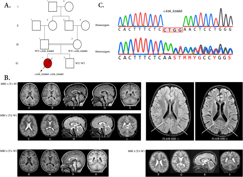
Fig. 1.
a) The pedigree of the family concerning the ADPRHL2 variant and the pertinent chromatograms. The parents were heterozygote for the frameshift deletion in the ADPRHL2 gene, while the patient (proband) was homozygote for this variant. The proband and her parents were subjected to whole-exome sequencing. In this pedigree, white symbols: unaffected who were homozygous for wild-type allele; red symbol: affected and homozygous for the variant; Square: male; circle: female; parallel lines: consanguineous marriage. Chromatograms showing nucleotide sequences of ADPRHL2 in the cDNA regions of c.636_639. The gap region in ‘homozygote’ indicates the deletion from C to G (4 nucleotides) in the patient. b) Serial brain MRI without contrast of the patient with an interval of two months (MRI 1 and 2) shows progressive cerebral and cerebellar atrophy in T1-W (A-E, K-O) and their cognate T2-W images (F-J, P-S) in horizontal, sagittal, and coronal planes (compare A, B with K, M for cerebral and C, D with N, R for cerebellar atrophy). Besides, thin white arrows in FLIAR-MRI 1 and 2 exhibit progressive posterior deep white matter signal changes in two serial FLAIR sequences

