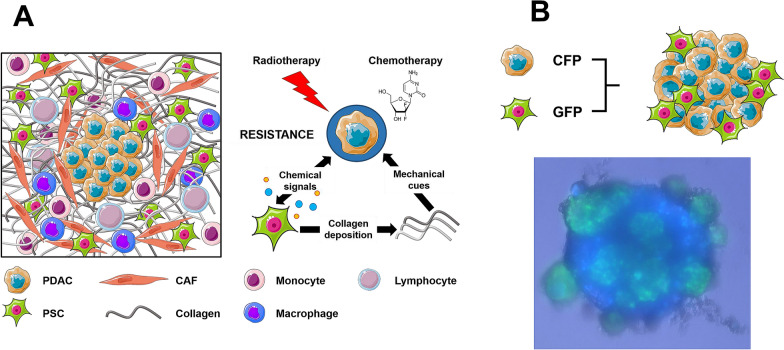
Fig. 1.
Schematic overview of the surrounding TME in pancreatic cancer. a The TME of PDAC is comprised of several components, including CAFs, immune cells and extracellular matrix proteins, like collagen. Activated PSCs and collagen are able to promote resistance against chemo- and radiation treatment via paracrine signaling and mechanical cues, respectively. b Representative fluorescent image of co-cultured PSC/PDAC (1:2) spheroid. Green fluorescent protein-expressing PSCs were co-cultured with cyan fluorescent protein-expressing primary PDAC cells. Image was taken 48 h post-seeding. CAF Cancer-associated Fibroblasts, PDAC Pancreatic Ductal Adenocarcinoma, PSC Pancreatic Stellate Cells, CFP Cyan Fluorescent Protein, GFP Green Fluorescent protein. Some material of this figure was adapted from images made by Servier Medical Art by Servier, licensed under a Creative Commons Attribution 3.0 Unported License, at https://smart.servier.com

