Fig. 2.
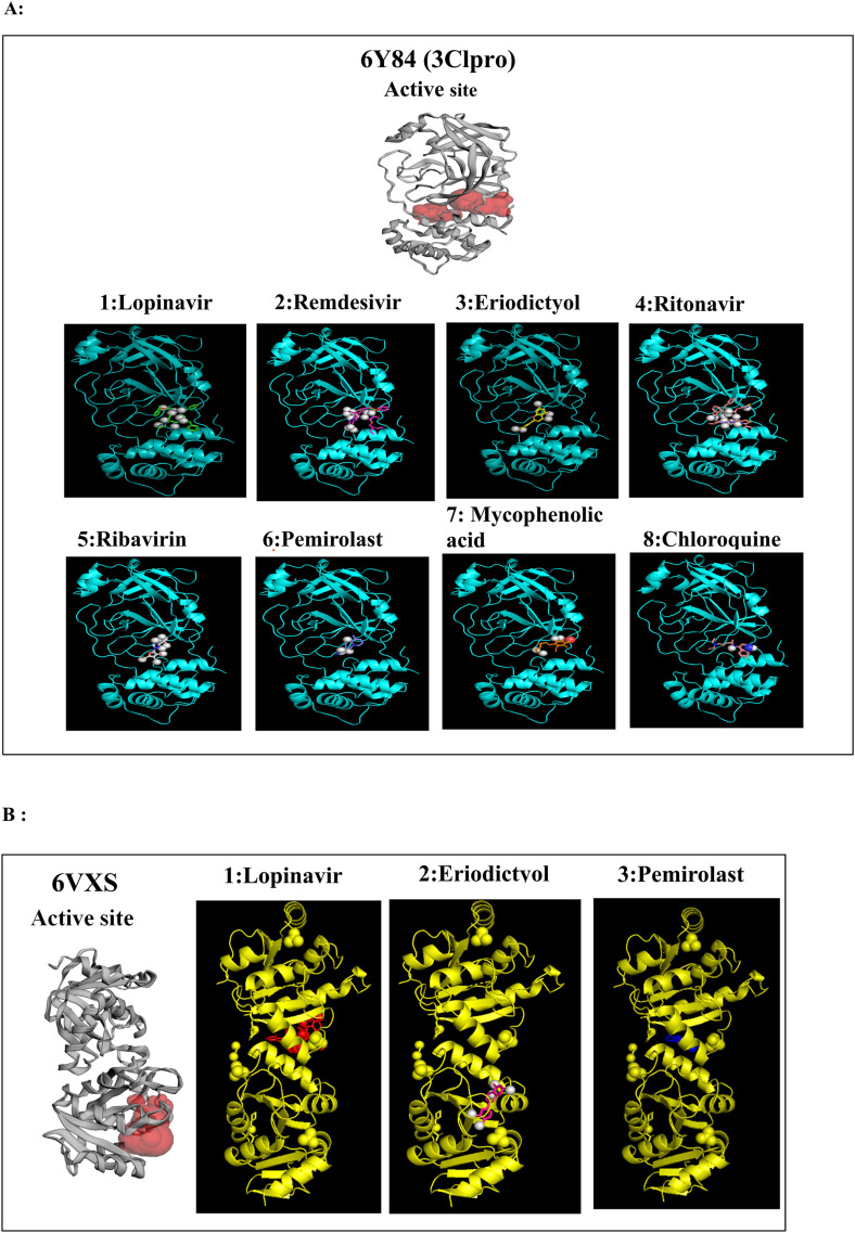
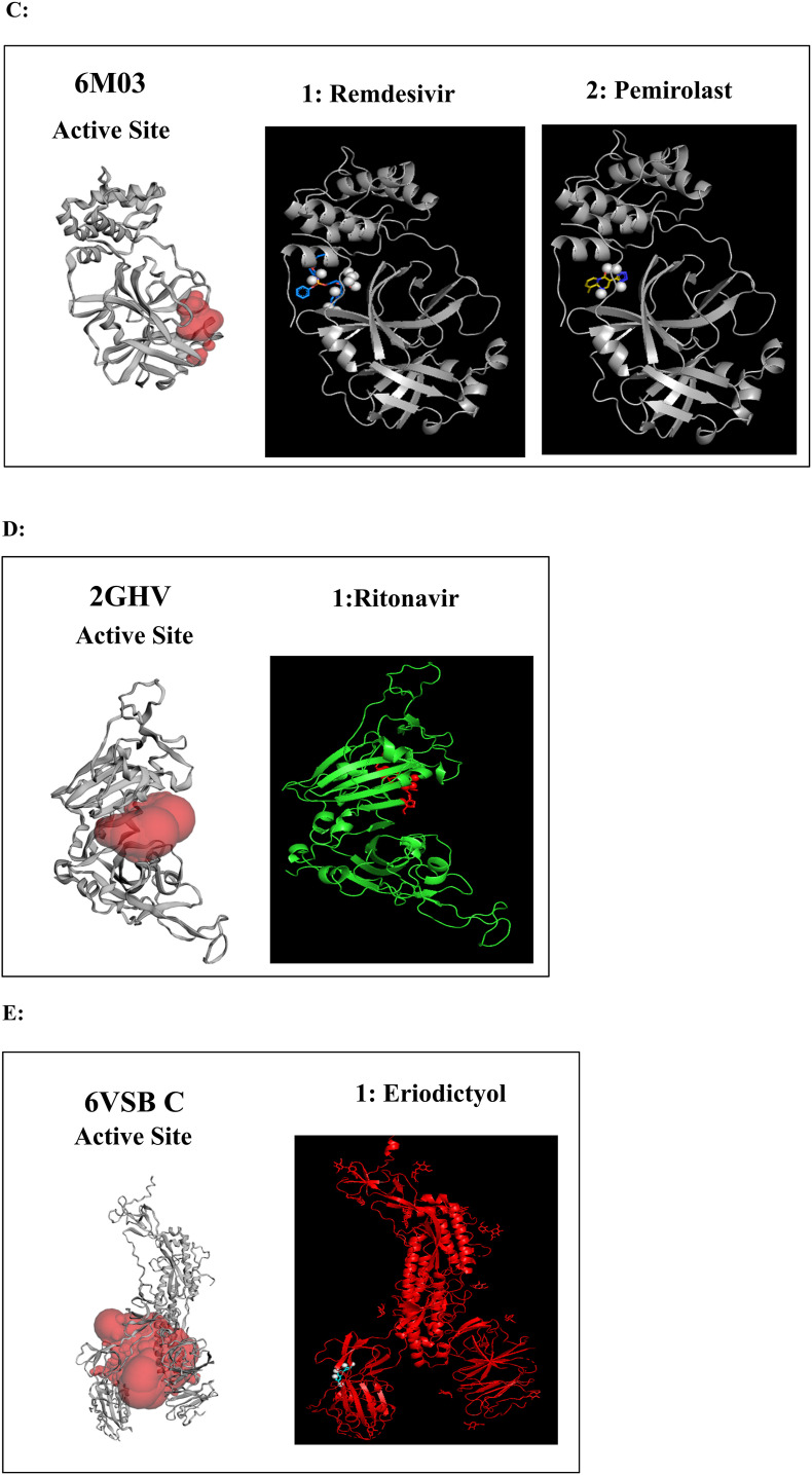
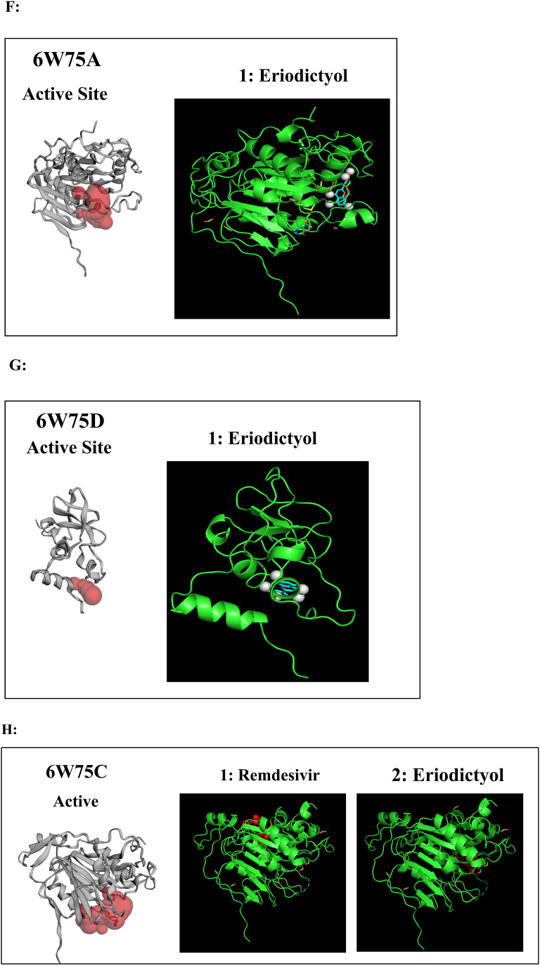
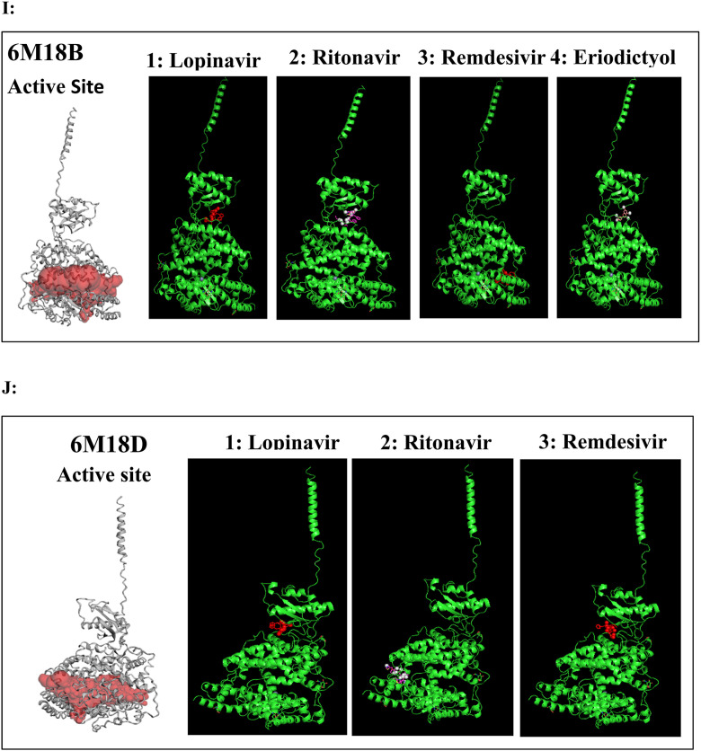
A to J: Represents the active site of a particular protein and binding poses of drug ligands showing binding energy less than -7.5 Kcal/mol. A: 3Clpro (6Y84) with 1: Lopinavir, 2: Remdesivir, 3: Eriodictyol, 4: Ritonavir, 5: Ribavirin, 6: Pemirolast, 7: Mycophenolic acid (MPA), 8: Chloroquine, B: Helicase (6VXS) with 1: Lopinavir, 2: Eriodictyol 3: Pemirolast, C: 3Clpro (6M03) with 1: Remdesivir, 2: Pemirolast, D: Receptro binding domain of S protein (2GHV) with 1: Ritonavir, E : S protein (VSB C chain) with 1: Eriodictyol, F: NsP10/16 complex (6W75A) with 1: Eriodictyol, G: NsP10/16 complex (6W75D) with 1: Eriodictyol, H: NsP10/16 complex (6W75C) with 1: Remdesivir, 2: Eriodictyol, I: ACE 2 receptor (6M18B) with 1: Lopinavir, 2: Ritonavir, 3: Remdesivir, 4: Eriodictyol, J: ACE 2 receptor (6M18D) with 1: Lopinavir, 2: Ritonavir, 3: Remdesivir.


