. 2020 Jul 31;2(4):e190053. doi: 10.1148/rycan.2020190053

Figure 5:

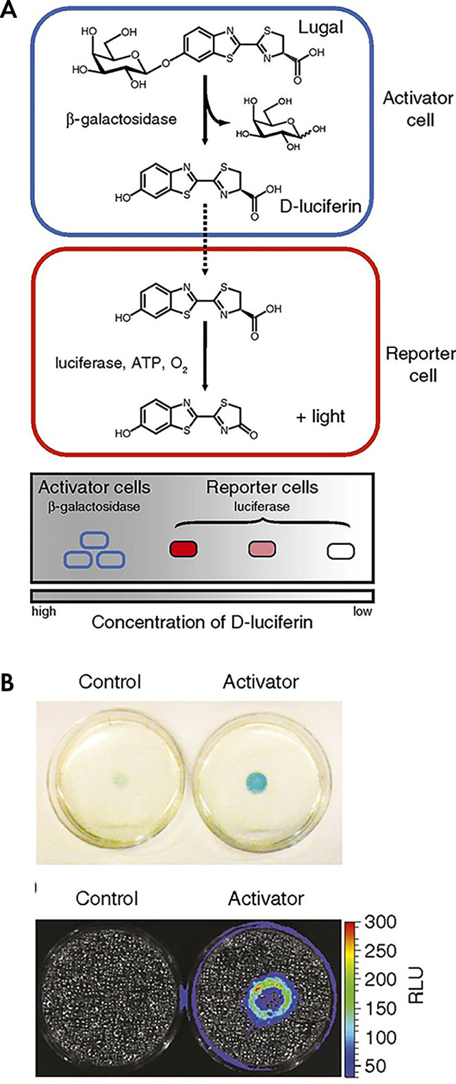
Dual enzyme–activated proximity sensor. A, Activator cells (expressing β-galactosidase) catalyze the cleavage of lugal, ultimately releasing d-luciferin. The liberated substrate enters nearby reporter cells, where it is used by luciferase to produce light. B, Reporter cells surrounding either control (left) or activator (right) cells were incubated with X-gal (5-bromo-4-chloro-3-indolyl-β-d-galactopyranoside) for 4 hours and imaged. The blue color correlates with β-gal activity. Representative bioluminescence image of cocultures after 48 hours of incubation and subsequent incubation with lugal. Each dish was incubated with lugal (100 μg/mL) for 1 hour before image acquisition. (Reprinted, with permission, from reference 53.) ATP = adenosine triphosphate, RLU = relative light units.

Dual enzyme–activated proximity sensor. A, Activator cells (expressing β-galactosidase) catalyze the cleavage of lugal, ultimately releasing d-luciferin. The liberated substrate enters nearby reporter cells, where it is used by luciferase to produce light. B, Reporter cells surrounding either control (left) or activator (right) cells were incubated with X-gal (5-bromo-4-chloro-3-indolyl-β-d-galactopyranoside) for 4 hours and imaged. The blue color correlates with β-gal activity. Representative bioluminescence image of cocultures after 48 hours of incubation and subsequent incubation with lugal. Each dish was incubated with lugal (100 μg/mL) for 1 hour before image acquisition. (Reprinted, with permission, from reference 53.) ATP = adenosine triphosphate, RLU = relative light units.