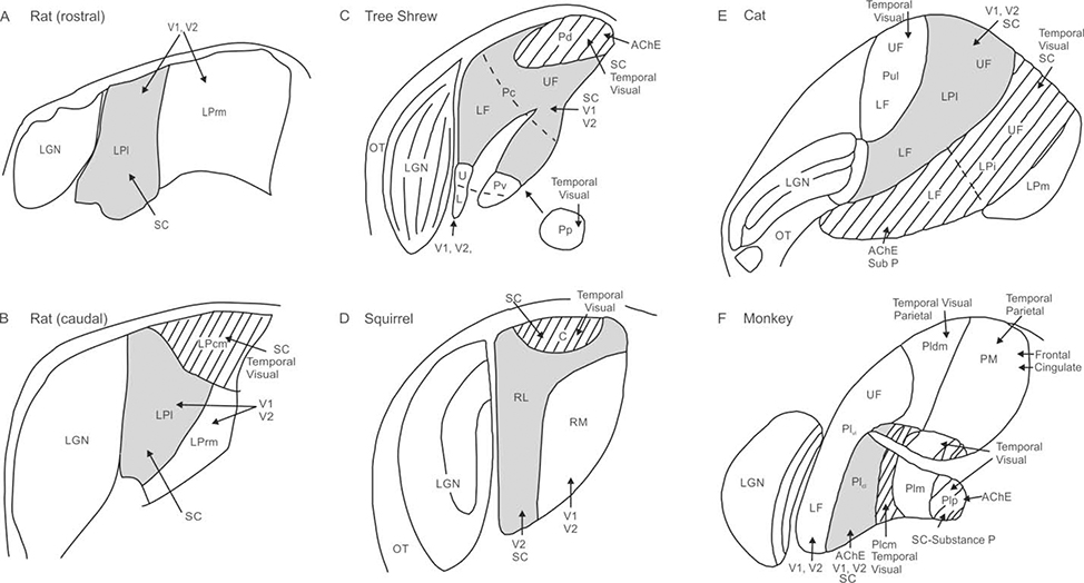
Figure 1.
Schematic of pulvinar anatomy across five species. Regions receiving dense superior colliculus (SC) input and with connections to temporal visual cortex (hatched) include caudomedial pulvinar (LPcm) in rat (B), dorsal pulvinar (Pd) in tree shrew (C), caudal pulvinar (C) in squirrel (D), intermediate LP (LPi) in cat (E), and inferior pulvinar (PI) in monkey (F), although not all of PI receives input from SC. Regions receiving sparse input from superior colliculus with connections to V1 and V2 (shaded grey) include lateral pulvinar (LPl) in rat (A, B), caudal pulvinar in the tree shrew (Pc; C), rostrolateral pulvinar (RL) in squirrel (D), lateral LP in cat (Lpl; E), and caudolateral inferior pulvinar (PIcl) in monkey (F). Regions with visual cortex connections only (no SC input) include rostromedial pulvinar (LPrm) in rat (A,B), ventral pulvinar (Pv) in tree shrew (C), rostromedial pulvinar (RM) in squirrel (D), lateral LP (LPl) in cat (E), and ventrolateral lateral pulvinar (PLvl) in monkey (F). Additional subregions include posterior pulvinar (Pp) in tree shrew, the pulvinar (Pul) and medial LP (LPm) in cat, and the medial (PM) and dorsomedial lateral (PLdm) pulvinar in monkey. Optic tract (OT) and lateral geniculate nucleus (LGN) are shown for reference. Adapted from Lyon et al. (2003) and Nakamura (2015); not to scale.

