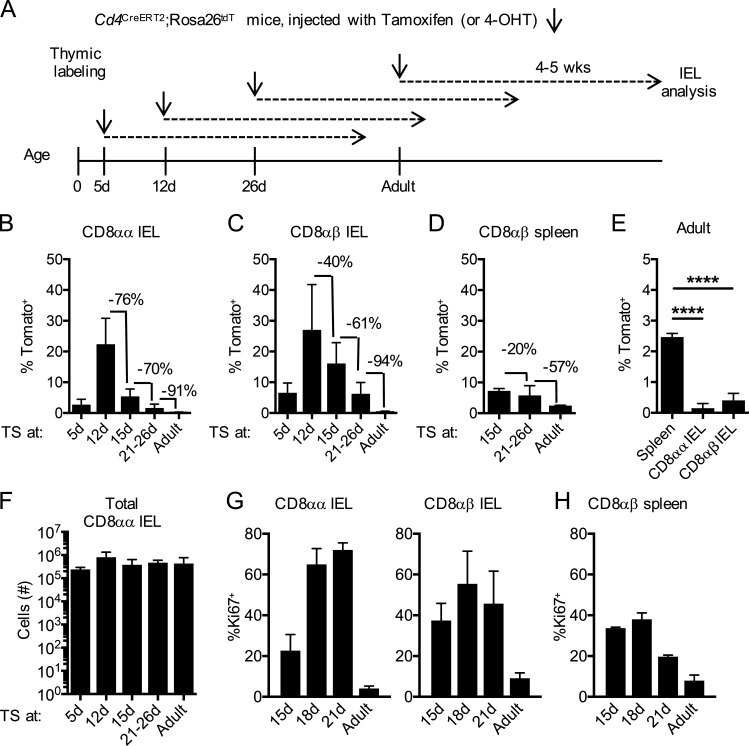
Figure 1.
Thymic IELps contribute to intestinal CD8αα IELs in early life. (A) Experimental outline: Cd4CreERT2×Rosa26tdT mice were time-stamped by intraperitoneal injection of tamoxifen (ages 21 d and older) or 4-OHT (ages 5–15 d). Tissues were analyzed 4–5 wk later by flow cytometry. (B–D) Frequency of Tomato-labeled cells within small intestinal TCRαβ+ CD8αα (B) or CD8αβ (C) IELs (n = 3 [5 d and 12 d]; n = 8 [15 d]; n = 6 [21–26 d and adult]) or CD8αβ+ T cells (D) in the spleen (n = 3 [15 d and adult]; n = 6 [21–26 d]) in animals time-stamped at the indicated ages. (E) Frequency of Tomato-labeled cells in the spleen (n = 3) or IELs (n = 6) of mice time-stamped as adults. (F) Numbers of CD8αα IELs in the small intestine at various ages (n = 3 [5 d and 12 d]; n = 8 [15 d]; n = 6 [21–26 d and adult]). (G) Frequency of Ki67+ cells within the CD8αα IELs and CD8αβ IELs at various ages (n = 5 [15 d]; n = 4 [18 d and 21 d]; n = 8 [adult]). (H) Frequency of Ki67+ cells within CD8αβ+ T cells in the spleen at various ages (n = 5 [15 d and 21 d]; n = 3 [18 d and adult]). Data in B–H are pooled from at least two independent experiments and show the mean ± SD. (E) ****P < 0.0001 (ANOVA with Bonferroni post-test). TS, time-stamped.

