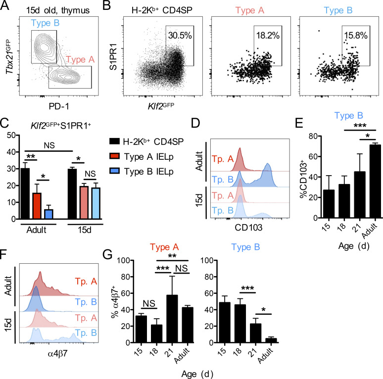
Figure 3.
Type B IELps have a thymus-emigrating phenotype in early life. (A) Flow cytometry for PD-1 and Tbx21GFP within CD25−CD1dtet−TCRβ+CD5+ DN thymocytes for the identification of type A (PD-1+Tbx21GFP−; red) and type B (PD-1−Tbx21GFP+; blue) IELps in 15-d-old Tbx21GFP mice. (B and C) Thymic H-2Kb+ (mature) CD4SP cells and type A (PD-1+; red) or type B (PD-1−; blue) IELps of 15-d-old Klf2GFP mice were analyzed for GFP and S1PR1 expression by flow cytometry (B), and the frequency of Klf2GFP+S1PR1+ cells was summarized (n = 3; C). (D–G) Type A (PD-1+Tbx21GFP−; red) and type B (PD-1−Tbx21GFP+; blue) IELps in 15- (n = 4–7), 18- (n = 8 or 9), or 21- (n = 3–5) day-old or adult (n = 3–5) Tbx21GFP mice were analyzed for surface expression of CD103 and α4β7. The expression levels in 15-d-old versus adult animals are shown as histograms (D and F), while the bar graphs depict the frequencies of CD103+ type B IELps (E) or α4β7+ type A and type B IELps (G). Data in C, E, and G are pooled from at least three independent experiments and show the mean ± SD. *P < 0.05, **P < 0.01, and ***P < 0.001 (ANOVA with Bonferroni post-test in C, E, and G). Tp., type.

