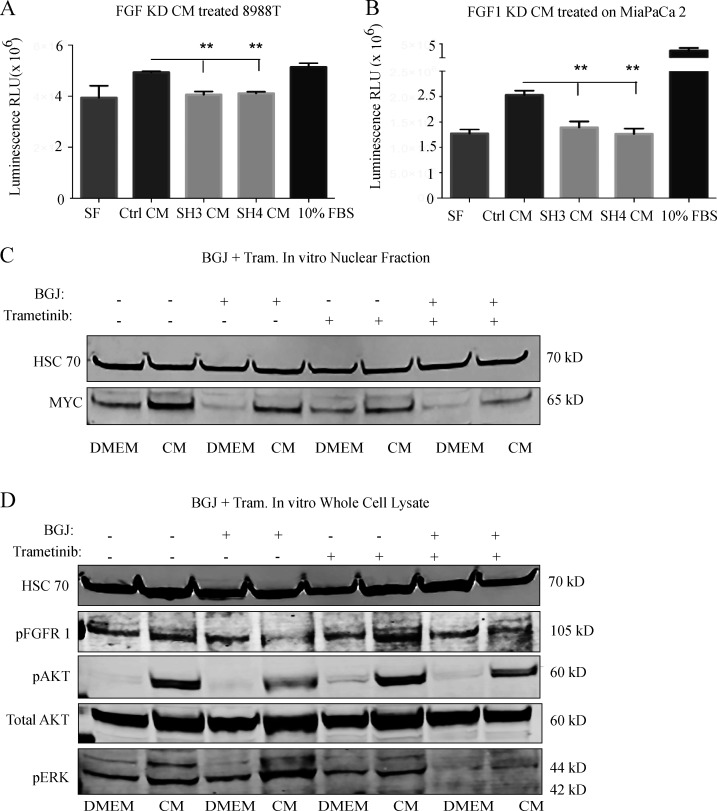
Figure S3.
The FGF1/FGFR axis augments MYC levels and PDAC growth in vitro. (A) Proliferation assay on 8988T cells treated with CM from control or FGF1 knockdown CAF 4414 for 72 h. **, P < 0.01 by one-way ANOVA (n = 3 independent experiments). (B) Proliferation assay for MiaPaCa2 cells treated with CM from control or FGF1 knockdown CAF 4414 for 72 h. **, P < 0.01 by one-way ANOVA (n = 3 independent experiments). (C and D) Western blots for (C) MYC and (D) the indicated signaling events in PSN1 cells treated with CAF 4414 CM, BGJ 398 (2 µM), and trametinib (20 nM) for 3 h.

