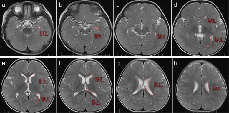
Fig. 2.
a–h Sketching ROI on lateral ventricle containing layer. The area of ROI is automatically revealed by workstation (a pons, b suprasellar cistern, c midbrain, d diaterma, e upper portion of the third ventricle, f diatela, g body of the lateral ventricle and posterior horn of the lateral ventricle, h upper portion of the lateral ventricles)

