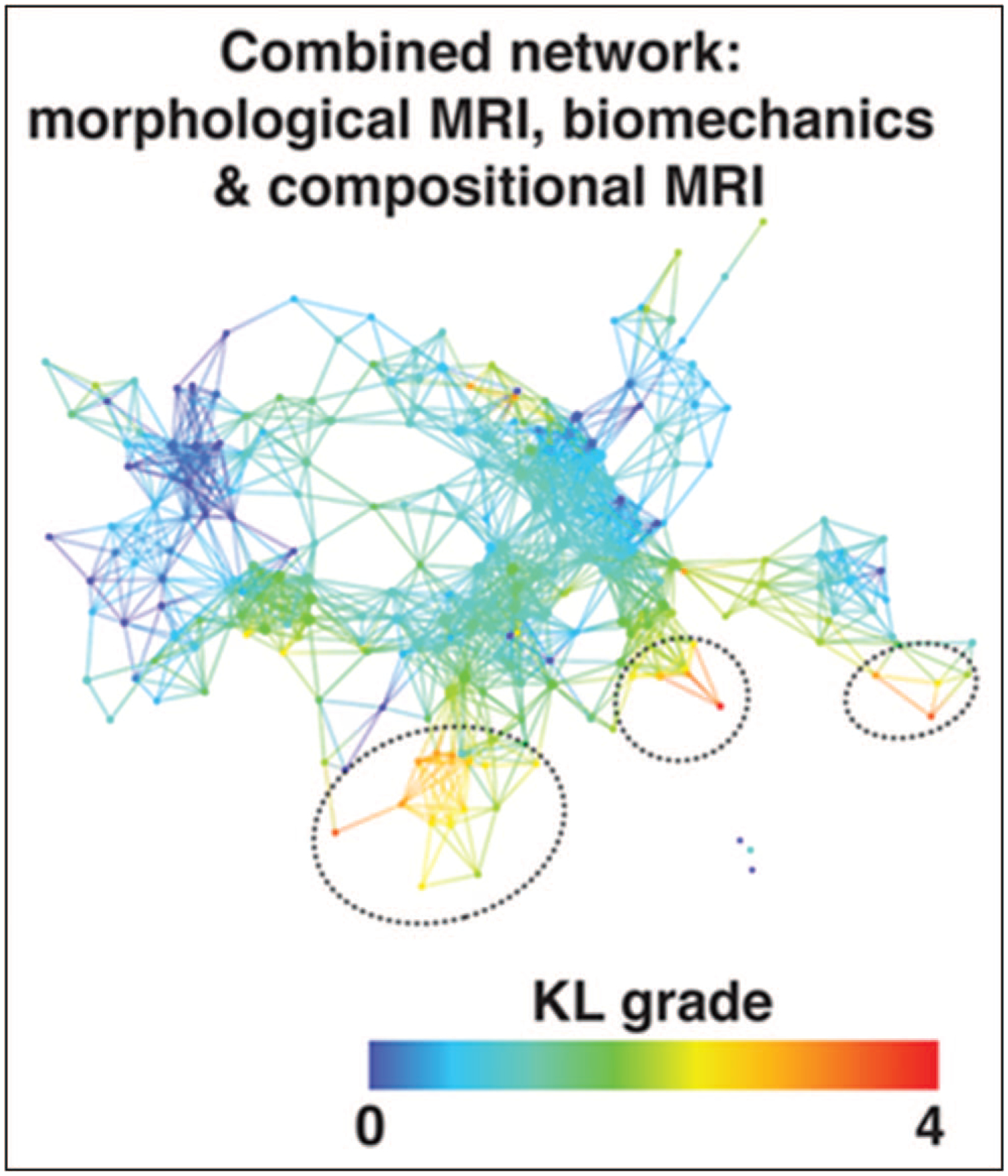
FIGURE 1.

Combined network of morphological MRI, biomechanics and compositional MRI. The network shows a gradient with severe patients appearing in the lower right (marked with dashed black circles) and less severe in the upper left based on Kellgren–Lawrence grading. Reproduced with permission [26■■].
