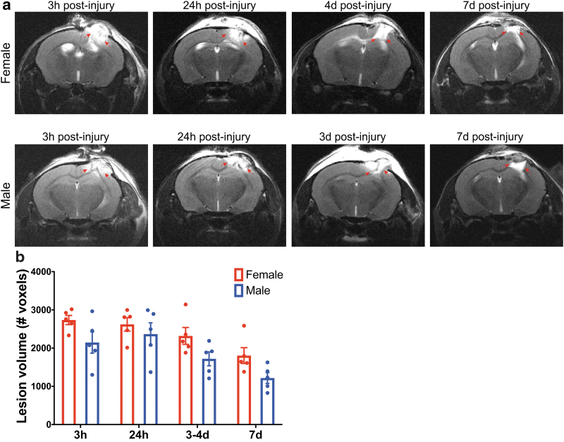
FIG. 4.
(a) Longitudinal in vivo T2-weighted MRI reveals changes in the cerebral cortex (right hemisphere) after CCI injury. A longitudinal cohort of female and male mice were repeatedly scanned at 3 h, 24 h, 3 days (male) or 4 days (female), and 7 days postinjury. Red arrows highlight the hyperintensity at and around the injury site. (b) Quantification of lesion volume T2-weighted scans acquired using a fast-spin echo sequence. Two-way ANOVA, post hoc Bonferroni's analysis of the lesion volume comparison between sexes showed no significant difference at 3 h, 24 h, 3–4 days, and 7 days postinjury. Mean ± SEM, n = 5. MRI, magnetic resonance imaging; SEM, standard error of the mean. Color images are available online.

