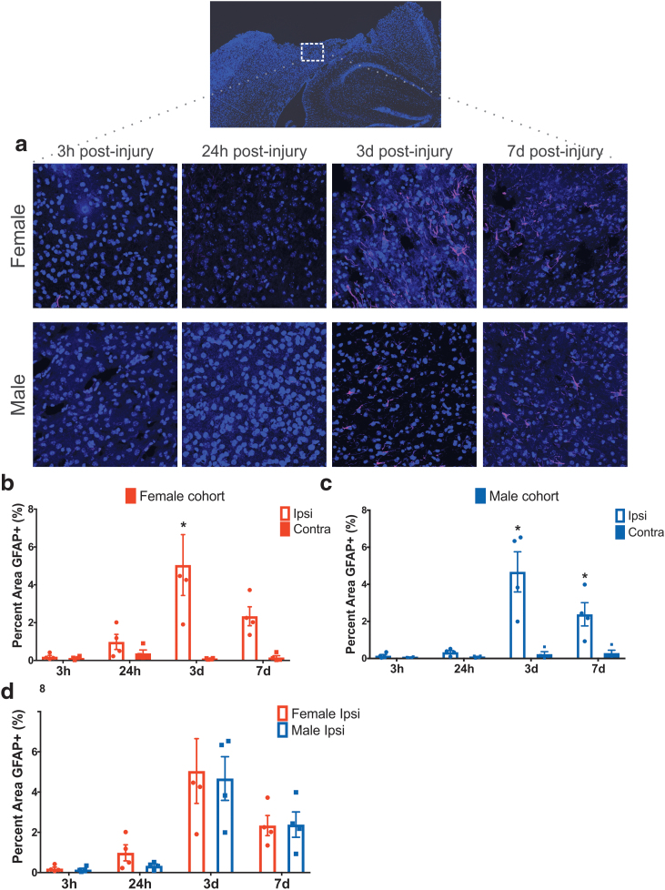
FIG. 5.
(a) Representative images of anti-GFAP staining within the cortical injury penumbra show progressive astrocyte reactivity at 3 h, 24 h, 3 days, and 7 days postinjury in both females and males. (b, c) Quantification of GFAP staining in the ipsilateral cortex in female cohort (b) and male cohort (c) at different time points postinjury. (d) Quantification of GFAP staining in the ipsilateral hemisphere of female and male cohorts across different time points, found no difference between sexes at 3 h, 24 h, 3 days, and 7 days postinjury. Two-way ANOVA mean ± SEM, n = 4 per group. GFAP, glial fibrillary acidic protein. Color images are available online.

