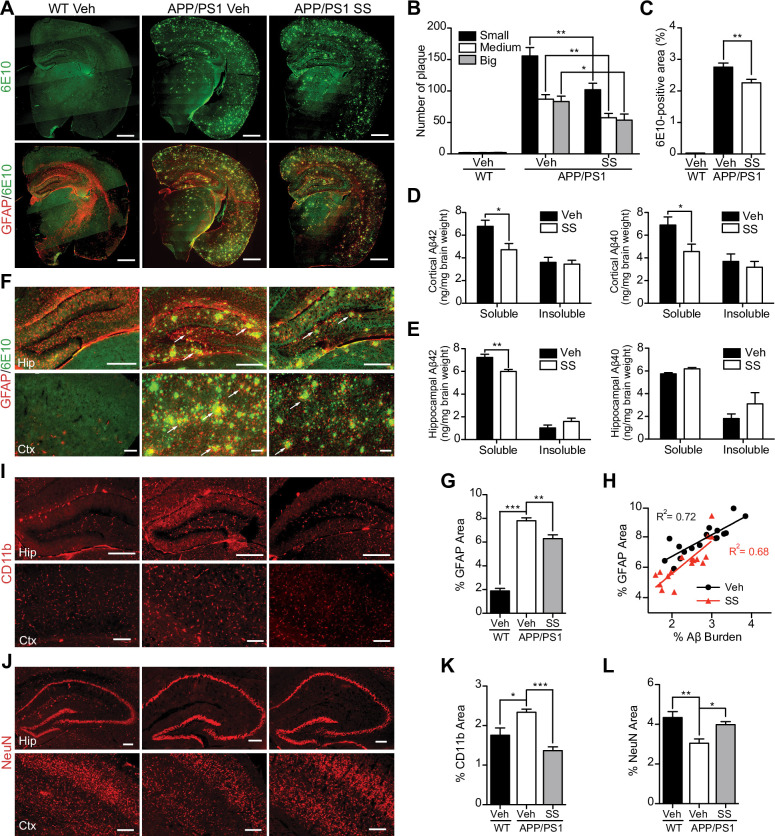
Fig 2. SS treatment alleviates Aβ levels and amyloid plaque burden, reduces gliosis and neuron loss in APP/PS1 mice.
(A–C) Representative half brain sections of WT mice, vehicle or SS-treated APP/PS1 mice stained with antibody against Aβ (6E10) and double staining of GFAP and 6E10 are shown. Scale bar, 1 mm. (B and C) Quantitative analysis of the number of 6E10-positive amyloid plaques (B) and Aβ covered area (C). n = 5 animals per group. (D and E) ELISA of soluble and insoluble Aβ40 and Aβ42 levels in cortical and hippocampal tissues of APP/PS1 mice. n = 6 for each group. (F, I and J) Representative images of WT mice, vehicle- and SS- treated APP/PS1 mice hippocampus and cortex double immunostaining of GFAP and 6E10 (F), CD11b (I) and NeuN (J). Arrows indicate astrocytes surrounding the amyloid plaques. Scale bar, 200 µm. (H) Coincidence of GFAP and Aβ burden in the brains of SS-treated APP/PS1 mice (red; n = 17) and vehicle-treated APP/PS1 mice (black; n = 17; P<0.0001). (G, K and L) The histograms depict the mean GFAP (G), CD11b (K), and NeuN (L) positive area ± S.E.M. in three groups. *P<0.05, **P<0.01, ***P<0.001.

