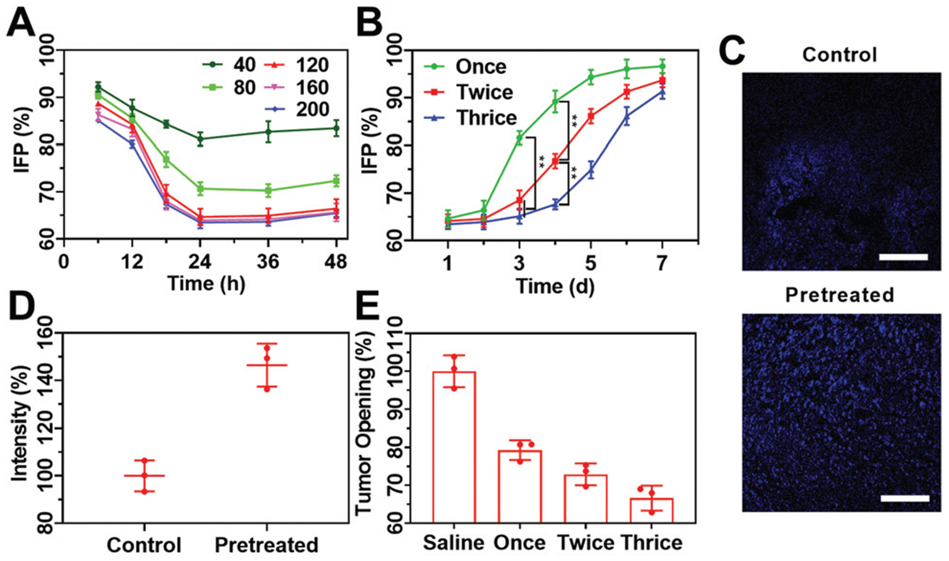
Fig. 5.

(A) Variation tendency of tumor IFP of H22 tumor-bearing mice with different injection doses (40, 80, 120, 160 and 200 mg kg−1) and interval times (6, 12, 18, 24, 36 and 48 h); (B) the tendency of tumor IFP during a week after receiving different treatments with a dose of 120 mg kg−1 and measured at 24 h after the last injection; (C) CLSM images of sectioned H22 tumor tissues stained with Hoechst 33342 with and without being pretreated with COP NPs thrice with an injection dose of 120 mg kg−1. Scale bar = 100 μm; (D) the average fluorescence intensity of Hoechst 33342 in three randomly selected equal areas from (C) quantified by ImageJ; (E) solid stress of CT26 tumors at 24 h after the injection of COP NPs at a dose of 120 mg kg−1. Data are represented as mean ± SD (N = 3).
