. 2020 Jul 23;9:e58571. doi: 10.7554/eLife.58571

Figure 7. Rad53-WT, but not Rad53-kd, can form a stable complex with DDK.

Figure 7.

(A) Gel-filtration analysis of purified Rad53-WT (top) or Rad53-kd (bottom), as indicated. Samples were analyzed by SDS-PAGE and Coomassie stain. (B) Gel-filtration analysis of Rad53-WT + DDK (top), DDK alone (center), or Rad53-kd + DDK (bottom). Samples were analyzed by SDS-PAGE and Coomassie stain or western blot, as indicated. (C) Model illustrating the inhibition of DDK-MCM DH complex formation by competitive binding of activated Rad53 to DDK.

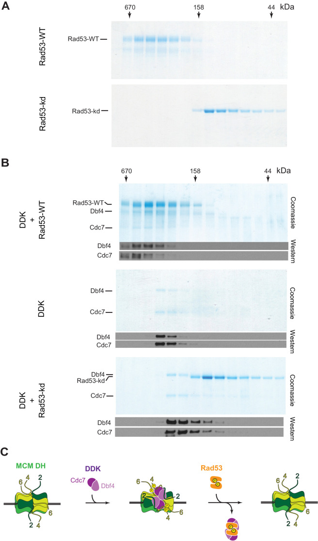
Figure 7—source data 1. Figure 7A, Rad53-WT.
Figure 7—source data 2. Figure 7A, Rad53-kd.
Figure 7—source data 3. Figure 7B, Rad53-WT + DDK.
Figure 7—source data 4. Figure 7B, Rad53-WT + DDK, immunoblot: Dbf4.
Figure 7—source data 5. Figure 7B, Rad53-WT + DDK, immunoblot: Cdc7.
Figure 7—source data 6. Figure 7B, DDK.
Figure 7—source data 7. Figure 7B, DDK, immunoblot: Dbf4, Cdc7.
Figure 7—source data 8. Figure 7B, Rad53-kd + DDK.
Figure 7—source data 9. Figure 7B, Rad53-kd + DDK, immunoblot: Dbf4, Cdc7.