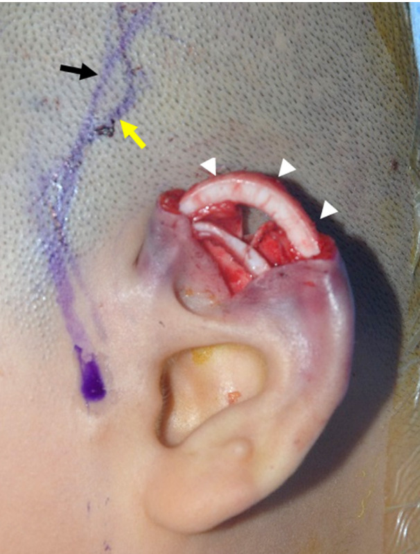
Fig. 4. Reconstructed upper helical framework.

The incision was designed after marking the superficial temporal artery (i.e., the pedicle of the superficial temporal fascial flap) using Doppler ultrasonography. The black arrow indicates the superficial temporal fascial flap (pedicle), while the yellow arrow indicates the incision line designed around the pedicle. The white arrowheads point to the reconstructed upper helical framework composed of cartilage from the eighth rib.
