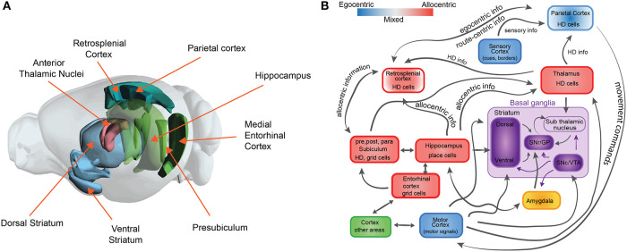
Figure 1.
The neuroscience of spatial navigation. (A) Key brain structures involved in rodent spatial navigation. Diagram adapted from the Allen Brain Atlas Explorer. (B) Schematic illustrating the general pattern of anatomical connectivity and the functional shift in frames of reference encoded by the brain regions that comprise the neural circuitry of spatial navigation. Hippocampus (HPC) and parahippocampal regions (entorhinal cortex, postsubiculum, and parasubiculum) encode an animal's position in space predominantly in allocentric or map-like coordinates. The blue and red boxes represent a spectrum denoting the relative density of egocentric (viewer-dependent, self-centered, or action centered frame of reference) vs. allocentric (map-like) encoding for each region. Retrosplenial cortex (RSC). Parietal cortex (PC) and anterior thalamic nucleus (ATN) are anatomically and functionally well-positioned to interface between egocentric and allocentric frames of reference within a larger navigational network. Purple boxes represent brain structures involved in value-based signals for conditional learning and spatial navigation (The basal ganglia circuit sub-diagram was inspired from Chersi and Burgess, 2015, used with permission).

