Fig. 1.

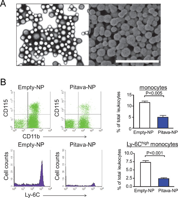
PLGA nanoparticles and their in vivo targeting of circulating inflammatory monocytes.
(A) PLGA nanoparticles prepared by the emulsion solvent diffusion method. Images were taken by the transmission electron microscope (left) and scanning electron microscope (right). Scale bar indicates 1 µm. (B) Flow cytometry of circulating leukocytes. CD11b+CD115+ cells are quantified as circulating monocytes (upper panels). Inflammatory monocytes determined by Ly-6C expression were evaluated (lower panels). Right graphs show quantitative data. N = 3–4.
