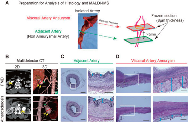
Fig. 1.

(A) Preparation for histological analysis and matrix-assisted desorption/ionization imaging mass spectrometry (MALDI-IMS). The arterial tissue was obtained from a patient who underwent aneurysmal resection and revascularization. We analyzed both the aneurysmal sac and adjacent artery, which is 5 mm from the aneurysm. (B) Computed Tomography (CT) revealed visceral artery aneurysms (VAAs), which cannot be distinguished from the fibromuscular dysplasia (FMD)-associated VAAs and atherosclerotic-VAAs. Two-dimensional (2D) (left) image and three-dimensional (3D) images of CT angiography (right). The yellow arrows indicate the aneurysm. Scale bar = 30 mm. (C)–(D). Elastica van Gieson staining of the adjacent artery (C) and VAA sac (D). Scale bar = 400 µm in each left panel. The area in the white box of each left panel is magnified in the corresponding panel on the right. Scale bar = 200 µm in each right panel.
