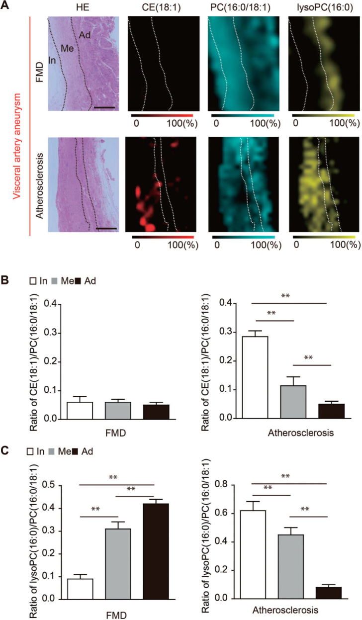
Fig. 2.

Analysis of the visceral artery aneurysm (VAA) using matrix-assisted desorption/ionization imaging mass spectrometry (MALDI-IMS).
(A) The distributions of choleterol ester (CE) (18:1), phosphatidylcholine (PC) (16:0/18:1), and lysophosphatidylcholine (lysoPC) (1-acyl16:0) in fibromuscular dysplasia (FMD)-associated VAA and atherosclerotic VAA are shown. Scale bar = 100 µm. HE, hematoxylin-eosin staining; In, intima; Me, media; Ad, adventitia (B) The ratios of CE (18:1) to PC (16:0/18:1) in the intima, media, and adventitia in FMD-associated VAAs and atherosclerotic VAAs. In, intima; Me, media; Ad, adventitia. **P < 0.01 indicates a statistically significant difference. (C) The ratios of lysoPC (1-acyl 16:0) to PC (16:0/18:1) in the intima, media, and adventitia in FMD-associated VAAs and atherosclerotic VAAs. In, intima; Me, media; Ad, adventitia. **P < 0.01 indicates a statistically significant difference.
