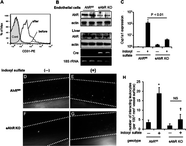
Fig. 1.

Effects of indoxyl sulfate on leukocyte-endothelial interactions in AhRfl/fl and eAhR KO mice. (A) Flow cytometry analysis of pulmonary cells stained with phycoerythrin-conjugated CD31 antibody before (dashed line) and after (line) endothelial cell isolation using CD31 microbeads. Gray filled-in line indicates cells stained with isotype control. (B) Immunoblotting images using anti-AhR antibody of isolated endothelial cells and liver from AhRfl/fl and eAhR KO mice. The mRNA expression of cre recombinase and 18S ribosomal (r)RNA was detected by reverse transcriptase-PCR. (C) AhRfl/fl and eAhR KO mice were subcutaneously injected with (+) 4 mmol/kg of indoxyl sulfate or left untreated (−) for 3 h, followed by isolation of endothelial cells from lung tissue. Cyp1a1 mRNA expression was determined by real-time PCR. (D, E, F, and G) Representative snapshots from intravital video microscopic analysis of leukocyte adhesive interactions in the femoral arteries (margins of vessels are indicated with dashed lines) of AhRfl/fl mice treated (D) with indoxyl sulfate or untreated (E) as well as eAhR KO mice treated (F) with indoxyl sulfate or untreated (G). White spots represent fluorescently labeled leukocytes visualized by intravenous injection of rhodamine 6G. (H) Quantitative analyses of leukocyte adhesion interactions in the femoral arteries. The data are expressed as means ± SEM (n = 6). *P < 0.05 versus (vs.) AhRfl/fl not treated with indoxyl sulfate, †P < 0.05 vs. AhRfl/fl treated with indoxyl sulfate.
