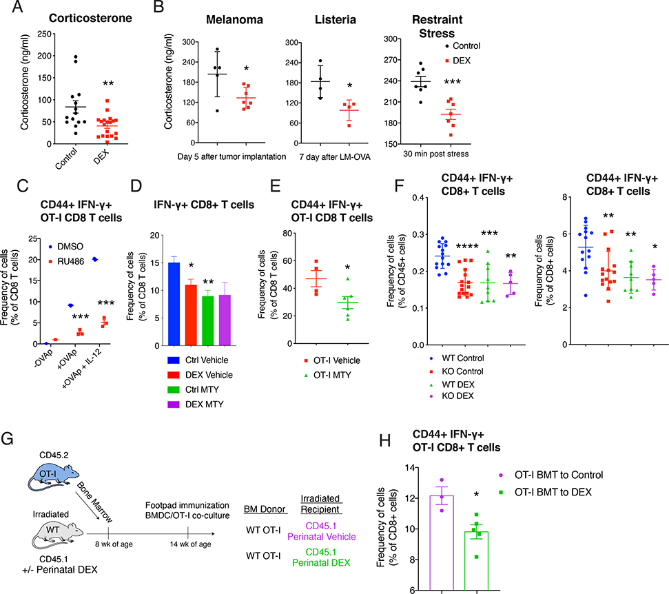
Figure 4. Perinatal GC exposure decreased systemic CORT level, and inhibition of GR signaling reduced CD8 T cell function.
(A) Corticosterone level in 16 week old B6 mice (n=14–19/group (female and male), ZT=3 (10 am)). (B) Corticosterone level in B6 mice after 5 days of B16-F10 melanoma implantation (n=5–7/group (male), left, ZT=4), after 7 days of LM-OVA (n=4/group (female), middle, ZT=4), and after 30 minutes of restraint stress (n=7/group (male and female), ZT=6). (C) Flow cytometric analysis of CD44+ IFN-γ+ OT-I CD8 T cells with or without RU486 (1 μg/ml) in BMDC and OT-I co-culture system described in Figure 1H (n=3/group (female), representative of 3 independent experiments including males). (D and E) MTY (800 μg/ml) was treated in drinking water during footpad immunization with LPS+IFA+OVA (−7 to +5 days of immunization) in mice, as described in Figure 1B. Flow cytometric analysis of IFN-γ+ CD8 T cells in the LN (16 week old Balb/c mice, n=4–7/group (male)) (D), and CD44+ IFN-γ+ OT-I CD8 T cells in the LN (16 week old OT-I mice, n=4–6/group (male)) (E). (F) 12 week-old Wild-type (WT;Nr3c1fl/fl) mice or KO (CD4-Cre Nr3c1fl/fl) mice were immunized with LPS+IFA+OVA. CD44+ IFN-γ+ CD8 T cells in LN were analyzed with flow cytometry (n=5–15/group (female and male)). (G and H) Bone marrow cells from 10 week old OT-I mice were transferred to 10 week old CD45.1+ wild-type mice. Footpads of recipient mice were immunized. Illustration of bone marrow transplantation experiment (G). Flow cytometric analysis of CD44+ IFN-γ+ CD8 T cells in LN (n=3–5/group (female)) (H). Data are represented as mean ± SEM. 1-way ANOVA (D and F) 2-way ANOVA (C), Student’s t-test (rest), *p<0.05, **p<0.01, ***p<0.001 vs Control (Vehicle, DMSO, WT Control) group.

