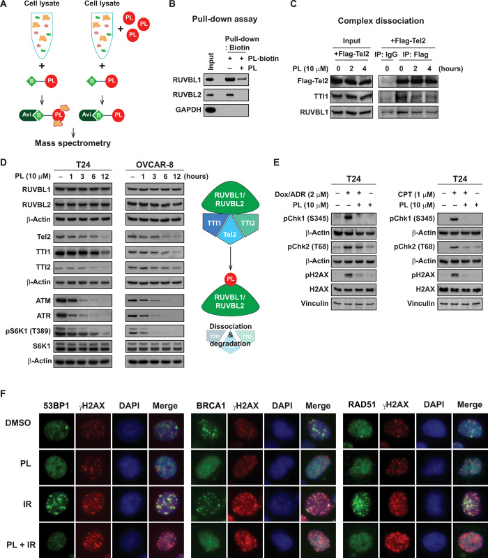
Fig. 3. PL targets the RUVBL1/2-TTT pathway.
(A) Target identification using mass spectrometry. Cell lysates were mixed with or without PL, and biotin-labeled PL was used to detect PL-binding proteins. Avidin beads were used to pull down PL-biotin adduct and was subjected to mass spectrometry analysis. (B) PL-biotin binds to RUVBL1 and RUVBL2 and free PL competes with this binding. Glyceraldehyde-3-phosphate dehydrogenase was used as a negative control. (C) The effect of PL on RUVBL1/2-TTT complex formation. T24 cells and T24 cells overexpressing Flag-Tel2 were used for immunoprecipitation with anti-Flag antibody and affinity agarose beads. PL (10 μM) was treated for 2 and 4 hours. (D) The effect of PL on RUVBL1/2-TTT proteins. T24 and OVCAR-8 cells were treated with PL (10 μM) for 1, 3, 6, and 12 hours and examined for RUVBL1/2-TTT proteins and its downstream PIKK pathway by immunoblotting. (E) PL suppresses doxorubicin (DOX)– and camptothecin (CPT)–induced DNA damage response signaling in T24 cells. PL was pretreated for 40 min before the addition of DOX (2 μM) and CPT (1 μM) for 1 hour. pChk1, pChk2, and pH2AX were examined by immunoblotting. (F) PL inhibits ionizing radiation (IR)–induced 53BP1, BRCA1, and Rad51 foci formation. PL was pretreated for 1 hour before IR at 4 gray for 3 hours.

