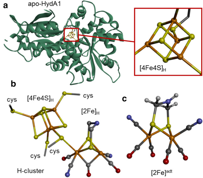
Fig. 1.

Crystal structures of iron components. Structures correspond to: a apo-HydA1 protein with the [4Fe–4S] cluster bound by four cysteines shown in magnification (PDB-ID 3LX4, ref [14]); b the complete H-cluster in the Hox state featuring a bridging CO ligand and an apical vacancy at the distal iron site of the diiron sub-complex (in a bacterial [FeFe]-hydrogenase, PDB-ID 4XDC, ref [15]); and c the synthetic diiron complex, [2Fe]adt, used for enzyme activation (color code: Fe, orange; O, red; N, blue; C, gray; H, white; protons were not resolved in protein structures; cys cysteine)
