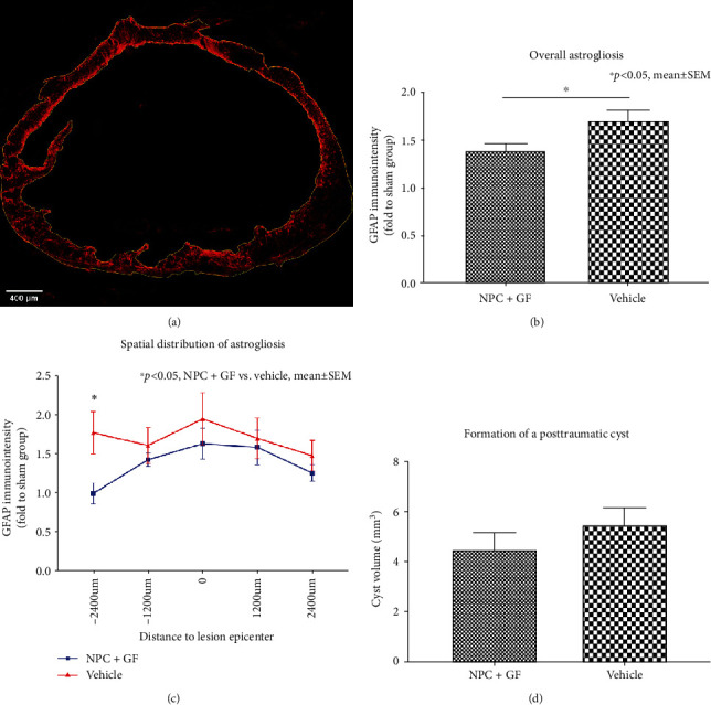
Figure 7.

In vivo astrogliosis and cyst formation 8 weeks after cervical SCI. (a) Spinal cord cross-section stained for GFAP (red) with ROIs (yellow) drawn around the entire spinal cord as well as the intramedullary, posttraumatic cyst (10x magnification). (b) Overall GFAP-immunointensity and (c) spatial distribution of GFAP-immunointensity expressed in proportion to (fold to) the sham group as a marker for reactive astrogliosis in the injured spinal cord. (d) Volume of the posttraumatic intramedullary cyst.
