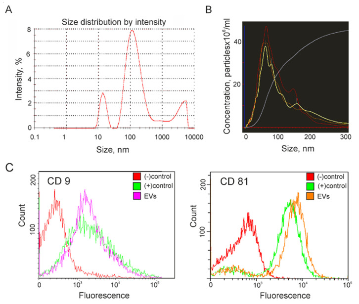
Figure 1.
Characterization of extracellular vesicles (EV) isolated from the conditioned culture medium (CCM) of Glia-Tr cell line. (A) The size of EV was quantified by dynamic light scattering (DLS). (B) Nanotracking particle analysis (NTA) of particle size and concentration. Each sample was measured in triplicate. (C) Flow cytometry analysis of isolated EVs for the surface expression of exosome markers CD9 (left panel) and CD81 (right panel). Immunobeads that were not incubated with EV during sample preparation were used as a negative control ((−) control). An aliquot of exosome standard (Lonza) was used as a positive control ((+) control).

