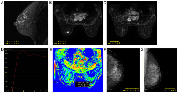
Figure 1.
Scans from a 50-year-old female patient. (A) A mass with irregular shape, (B) spiculation and (C) heterogeneous enhancement was observed in the left upper quadrant of the breast by enhanced magnetic resonance imaging. (D) The early enhancement rate was 20.3%, and the time-intensity curve was of type III. (E) The observed diffusion coefficient value was 0.849×10⁻³ mm2/sec. The invasive micropapillary carcinoma was surrounded by a non-mass enhancement lesion with segmental distribution and an internal cluster-like enhanced region, which was pathologically confirmed as simultaneous ductal carcinoma in situ. (F and G) Mammography revealed a slightly dense mass with an irregular shape and spiculation.

