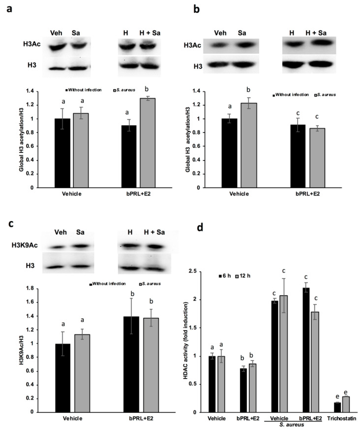
Figure 3.
Regulation of H3 acetylation by bPRL and E2 in bMECs during infection. Densitometrical analysis of the immunoblots that shows the relative expression of H3Ac with respect total H3 in bMECs treated for 12 h (a) or 24 h (b) with the combined hormones. Additionally, representative western blot analysis is shown. In (c), the densitometrical analysis of the immunoblots that shows the relative expression of H3K9Ac with respect total H3 in bMECs treated for 12 h with the combined hormones is shown with its respective representative western blot analysis. Each bar shows the mean ± SE of optical density (arbitrary units, AU), considering the expression of control cells (1% ethanol) as 1 (data normalized), from four different experiments (n = 4). (d) bMEC lysates were obtained at 6 or 12 h of treatment with combined hormones with or without 2 h of S. aureus infection, and were incubated with the substrate for class I and II histone deacetylases (HDACs) accordingly to the manufacturer’s instructions. Total HDAC enzyme activity was determined by using the HDAC fluorometric cellular activity assay Fluor de Lys. Each bar shows the mean of HDAC activity of cells from two different experiments ± SE, which were run in duplicate (n = 4), considering the expression of control cells (1% ethanol) as 1 (data normalized). Different letters indicate significant differences between each condition (one-way ANOVA, post hoc Tukey test p ≤ 0.05). Trichostatin (1 μM) was employed as an inhibitor of HDACs. Veh: vehicle; Sa: S. aureus; H: combined hormones.

