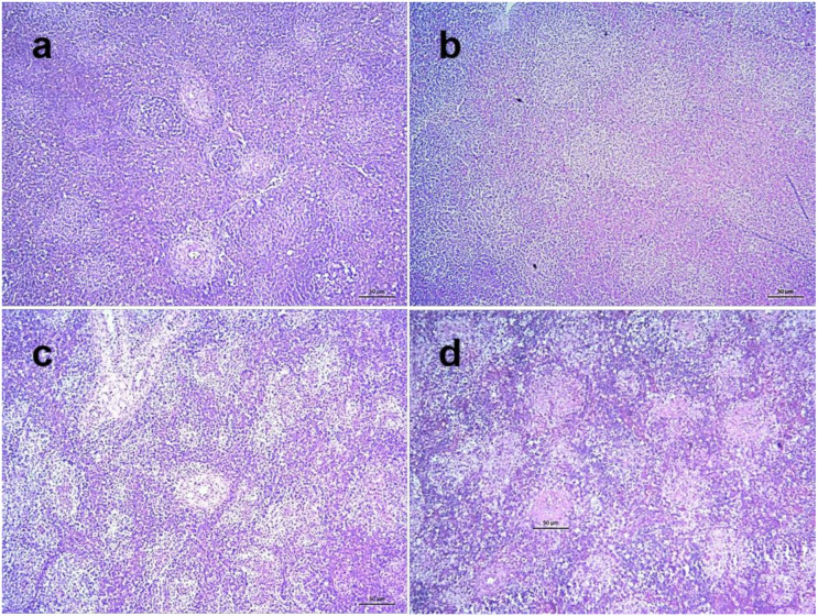
Figure 9.
Histopathological changes in the spleen of NDV infected chickens. Section of the spleen of control chickens showing normal histology (a). Sections of spleens of NDV infected chickens showing normal architecture at 2 dpi (b), mild multifocal lymphoid depletion in the cortex of spleen at 3 dpi (c) and severe multifocal lymphoid depletion with increased number and sizes at 5 dpi (d). H&E stain, bar indicates magnification.

