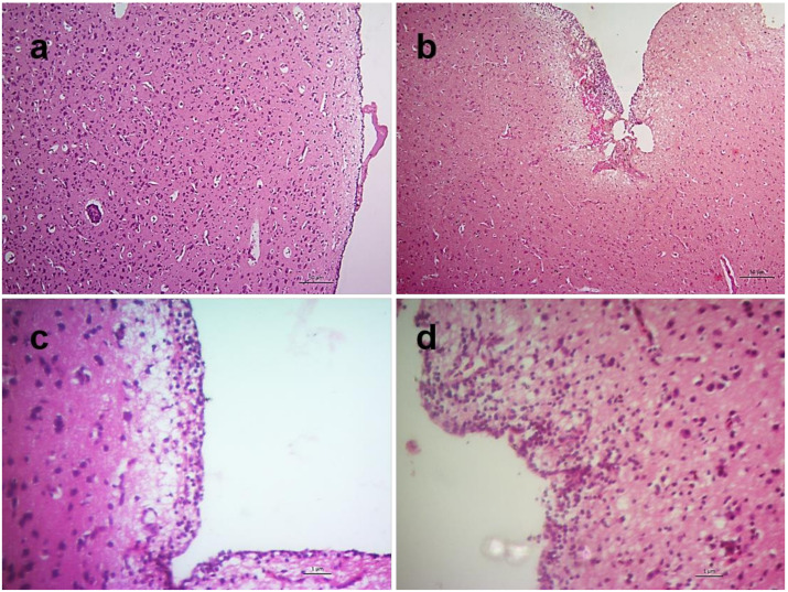
Figure 11.
Histopathological changes in the brain of NDV infected chickens. Section of the brain of control chickens showing normal architecture (a). Sections of brains of infected chickens showing focal meningitis at 2 dpi (b), diffuse meningitis with encephalomalacia at 3 dpi (c) and diffuse meningoencephalitis with encephalomalacia at 5 dpi (d). H&E stain, bar indicates magnification.

