Figure 5.
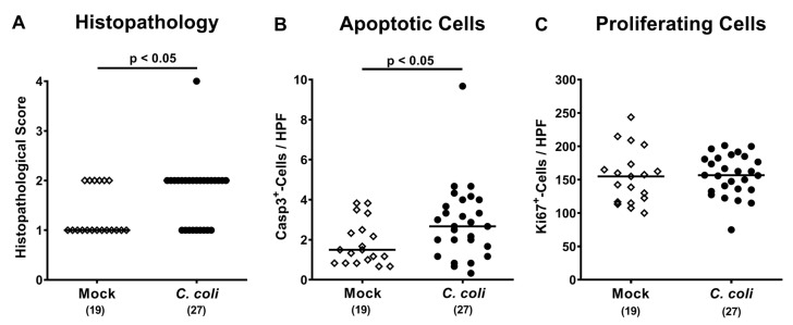
Microscopic inflammatory responses in the colon following peroral C. coli infection of aged conventional IL-10-/- mice. Aged IL-10-/- mice were perorally challenged with C. coli (black circles) on days 0 and 1 or received vehicle (mock controls; white diamonds). Upon necropsy (i.e., day 28 post-infection), (A) colonic histopathological changes were quantitatively assessed in hematoxylin and eosin (H&E) stained paraffin sections applying a histopathological scoring system (see methods). Additionally, the average numbers of colonic epithelial (B) apoptotic (cleaved caspase 3+; Casp3+) and (C) proliferating (Ki67+) cells were assessed microscopically from six high power fields (HPF, 400 x magnification) per animal in immunohistochemically stained large intestinal paraffin sections. Medians (black bars), levels of significance (p-values; assessed by the Mann–Whitney U test and Student’s t test) and numbers of analyzed mice (in parentheses) are indicated. Data were pooled from four independent experiments.

