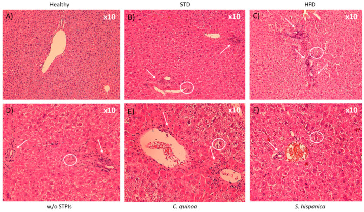
Figure 2.
Liver hematoxylin and eosin staining of 14 weeks-old high fat diet-fed C57BL/6 mice administered with or without PIs (100 µg/animal) (A,B). At the end of the study period in the w/o STPIs group, 4 animals were analyzed, while in those administered with C. quinoa and S. hispanica 6 animals were analyzed. Arrows indicate prominent accumulation of nodes of mononuclear cells after 14 weeks. Circles locate irregular contours of the bile duct with nuclear pseudostratification and vacuolization of the epithelium, with marked portal inflammation (C,D). (E,F) Artifice that produces steatosis suggestive image.

