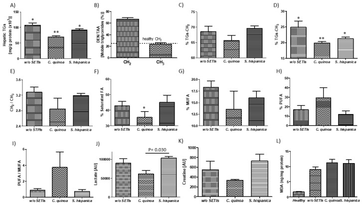
Figure 3.
1H-MAS MR analyses of DEN/TAA-treated mice fed high fat diet administered with or without protease (serine-type) inhibitors (PIs) (100 µg/animal). (A) Colorimetric determination of total hepatic triglyceride content. 1H-MAS MR levels of triglyceride-CH2 (B) and triglyceride-CH3 (C) in tissue from HCC developing mice. (D–F) Lipid patterns for saturated fatty acids, MUFA and PUFA. (G–I) 1H-MAS MR levels of lactate (J), creatinine (K). Quantification of the oxidation marker malonaldehyde (MDA) in liver samples of HCC developing mice (L). Results are expressed as mean ± SEM (n = 4−6). * Indicates statistical differences (Tukey–Kramer’s test, p < 0.05) to DEN/TAA-treated animals not receiving PIs.

