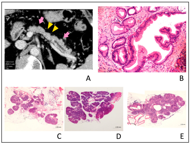
Figure 6.
Typical computed tomography (CT) images showing partial pancreatic parenchymal atrophy (PPA) in an 81-year-old man with stage 0 (carcinoma in situ) pancreatic cancer. Localized main pancreatic duct (MPD) stenosis without tumor lesion in the pancreatic body was detected in CT for further examination of a small cyst in the pancreatic tail. (A); The area exhibiting PPA had an atrophic change corresponding to the distribution of MPD stenosis (yellow arrow head) and defined as localized atrophy compared with the upstream and downstream parenchyma (pink arrow). (B); Hematoxylin and eosin (H&E)-stained resected specimen. Histopathological examination showed high-grade pancreatic intraepithelial neoplasia (PanIN) of the main pancreatic duct (H&E stain, ×20 magnification). (C); Severe atrophy and fibrosis of the pancreatic parenchyma and focal fatty change adjacent to the high-grade PanIN (H&E stain, ×0.5 magnification). (D); Downstream side of pancreatic parenchyma had no atrophic change as compared with Figure 6C (H&E stain, ×0.5 magnification). (E); Upstream side of pancreatic parenchyma had slight fibrosis and focal fatty change as compared with Figure 6C. Acinar cell architecture was maintained (H&E stain, ×1 magnification).

