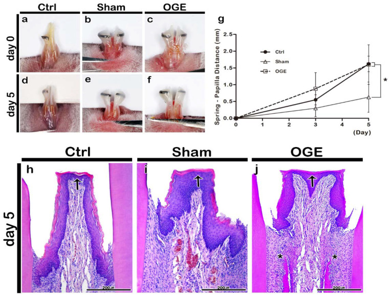
Figure 2.
Morphological and histological changes of interdental papilla (IDP) after the wire attachment. Morphology of IDP in control (a,d), sham (b,e), and open gingival embrasure (OGE) (c,f) groups on days 0 (a–c) and 5 (d–f) post-wire attachment. Change in spring-papilla distance (SPD) value during the wire attachment period (g). The SPD values in OGE and control groups were significantly higher than those in sham group (p < 0.0167). Histological analysis of IDP in the control (h), sham (i), and OGE (j) groups after wire attachment. Arrows and asterisks indicate the IDP and high cell density around the alveolar bone, respectively. * p-value was obtained from Kruskal–Wallis test, followed by Bonferroni’s post hoc test (p < 0.0167). Scale bars = 200 µm (h–j).

