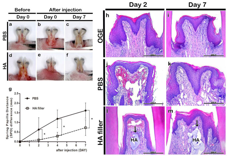
Figure 3.
Morphological and histological changes in interdental papilla (IDP) after phosphate buffer saline (PBS) or hyaluronic acid (HA) filler injection. Morphology of IDP in the open gingival embrasure (OGE) group before (a,d) and immediately after injection (b,e), and on day 7 (c,f) post-PBS (a–c) or -HA filler (d–f) injection. The spring-papilla distance (SPD) values of the PBS injection group were higher than those of the HA filler injection group on days 2, 4, and 7 post-injection (g) (p < 0.05). Histological analysis of IDP on days 2 (h,j,l) and 7 (i,k,m) post-injection. Arrows indicate HA filler. HA, hyaluronic acid. * p-value was obtained from Mann–Whitney U test (p < 0.05). Scale bars = 200 µm (h–m).

