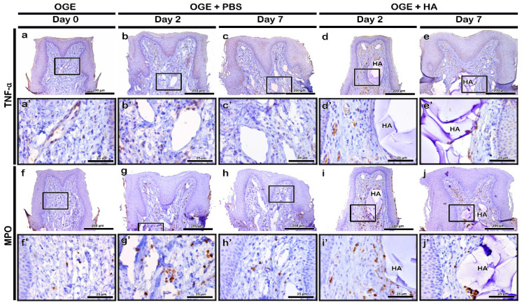
Figure 5.
Immunolocalization of tumor necrosis factor (TNF)-α and myeloperoxidase (MPO) in interdental papilla (IDP) after phosphate buffer saline (PBS) or hyaluronic acid (HA) filler injection. The localization patterns of TNF-α (a–e) and MPO (f–j) in IDP on days 2 and 7 post-PBS (b,c,g,h) or HA filler (d,e,i,j) injection. Higher magnification view of box in a–j (a’–j’). Scale bars: 200 µm (a–j) and 25 µm (a’–j’).

