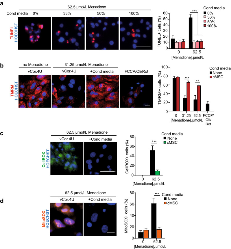
Figure 3.
Mouse cMSCs’ secretome blocks human ventricular myocyte apoptosis and the dissipation of mitochondrial potential induced by menadione. (a) Representative images and bar graph of TUNEL+ vCor.4U human ventricular myocytes, 24 h after menadione ± cMSC-conditioned media at the concentrations shown. n = 9. (b) Representative images and bar graph of mitochondrial TMRM in vCor.4U hPSC-CMs, stressed with menadione ± cMSC-conditioned media. Carbonyl cyanide 4-(trifluoromethoxy) phenylhydrazone (FCCP), oligomycin (Oli) and rotenone (Rot), uncouplers of oxidative phosphorylation, were used as controls. n = 7. (c) Representative images and bar graph of CellROX in vCor.4U hPSC-CMs 8 h after menadione ± cMSC-conditioned media. n = 12. (d) Representative images and bar graph of MitoSOX 4 h after menadione ± cMSC-conditioned media. n = 7. For all panels: scale bar 50 μm; data are shown as the mean ± SEM; *p < 0.05; ***p < 0.0001.

